The Sternoclavicular Joint
Anatomy of the Sternoclavicular Joint
The sternoclavicular joint (SCJ) is a unique joint within the body. It is the only connection between the upper limb and the rest of the skeleton, and as a result, has the strongest stabilizing ligaments in the body. It is a saddle-shaped joint and, like the knee, has a cartilage between its joint surfaces. Fortunately, because of its central position, the SCJ is rarely face Sternoclavicular Cambridge injuries.However, when an injury does occur it can cause a serious problem. Whilst Sternoclavicular Cambridge injuries to the SCJ are rare osteoarthritis (wear-and-tear arthritis) is very common athough it is often asymptomatic.
A knowledge of the normal anatomy of the shoulder and its complex arrangement of muscles, ligaments and tendons helps us to understand why a shoulder problem might occur and how to treat it.
- The SCJ is a synovial joint formed between the clavicle (collar bone) and the sternum (chest bone)
- The size of the articular surface on the sternal side of the joint is very small when compared to the clavicular side, potentially making the joint unstable
- The SCJ is very rarely dislocated due to an extensive group of very strong ligaments that surround the joint
- The SCJ is further stabilized by various muscle groups that are attached around it

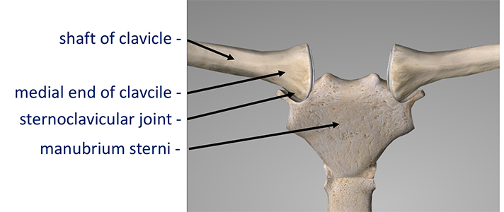
Bones of the SCJ
- The SCJ is formed by the medial (inner) end of the clavicle and a small facet on the top edge of the sternum
- The medial end of the clavicle is a relatively bulbous and relatively large, in comparison with the Sternal side of the joint. Only the lower two-thirds of the clavicle is covered with articular cartilage. The capsule and articular disk have a broad insertion into the top third
- Although only a small amount of movement occurs at the joint it is able to move in 3-axes (up and down, backwards and forwards and rotation). This movement is essential for the rest of the ‘shoulder girdle’ to work properly


SCJ Stabilising Ligaments
The SCJ is stabilised by a surrounding group of extremely strong ligaments. The Sternoclavicular Ligaments are not ‘true’ ligaments but are a confluent thickening of thejoint capsule. The Costocalvicular Ligaments run from the first rib to the medial end of the clavicle and the Interclavicular Ligament runs between the top of the SCJs. If you are suffering from a Sternoclavicular Cambridge injury, there’s no need to worry as you can always rely on Cambridge Shoulder to provide you with the best treatment for your Sternoclavicular Cambridge injury.
- Anterior Sternoclavicular Ligament – this is an oblique capsular thickening at the front of the joint. It also forms the anterior attachment of the intra-articular SCJ disk and stabilizes the joint in the anterior – posterior (front – back) plane.
- Posterior Sternoclavicular Ligament – this is a broad thickening of the posterior capsule and stabilizes the joint in the anterior – posterior (front – back) plane.
- Interclavicular Ligament – This is a clearly defined ligament that runs between the two clavicular heads. It is involved in stabilizing the SCJs in the superor plane
- The Costoclavicular Ligament – this is a short and very robust ligament that runs from the first rib and costal cartilage onto the medial end of the clavicle. It is an important joint stabilizer
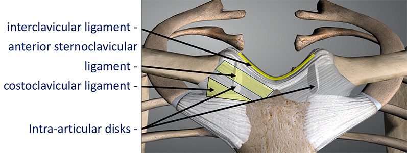
SCJ Intra-Articular Disk
TThe Rotator Cuff tendons are the next layer around the shoulder joint. A tendon is the connection of a muscle onto a bone.
- The Rotator Cuff Tendons – this is formed by a group of 4 tendons that connect the deepest layer of 4 muscles to the humerus. These muscles are,
- - Subscapularis
- - Supraspinatus
- - Infraspinatus
- - Teres Minor
The Intra-Articular disk is a strong fibrocartilaginous disk that runs through the centre of the joint and plays a role in load dissipation and joint stability.
- The complete Intra-Articular Disk divides to joint into clavicular and sternal sides
- Like the menisci (cartilages) in the knee the Intra-Articular Disk is formed from fibrocartilage and, like the menisci in the knee, is vulnerable to tearing
- The Intra-Articular Disk has a very broad attachment to the top third of the clavicular head and inserts circumferentially into the anterior, inferior and posterior capsule
- The Intra-Articular disk plays a role in stabilizing the joint and in dissipating some of the load that passes across the joint
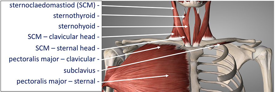
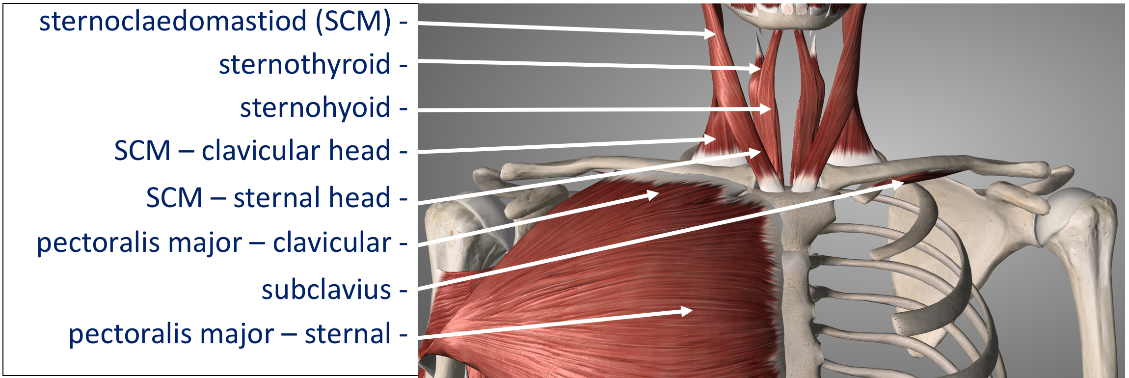
SCJ Stabilising Muscles
Like the Glenohumeral joint (Shoulder Joint) the SCJ has a group of surrounding muscles which help to stabilise the joint. The various muscles work synergistically to add an additional ‘dynamic envelope’ of stability to the SCJ
- Subclavius – this muscle has a long origin from the top of the first rib inserting on to the underside of the medial clavicle. It helps to depress the joint
- Sternocleidomsatoid (SCM) – this is a very important muscle that arises from the base of the skull and splits into 2 heads. The Sternal Head inserts into the top of the Manubrium and the Clavicular Head inserts onto the medial end of the clavicle. It plays an important role in anterio – posterior (front – back) stability
- Pectoralis Major – the clavicular insertion of the big Pectoralois Muscle helps to depress the SCJ and prevent superior instability
SCJ - Conditions & Injuries
- Arthritis of the Sternoclavicular Joint
- Instability of the Sternoclavicular Joint
- Sternoclavicular Joint Surgery