XRays
X-Rays for Shoulder Problems
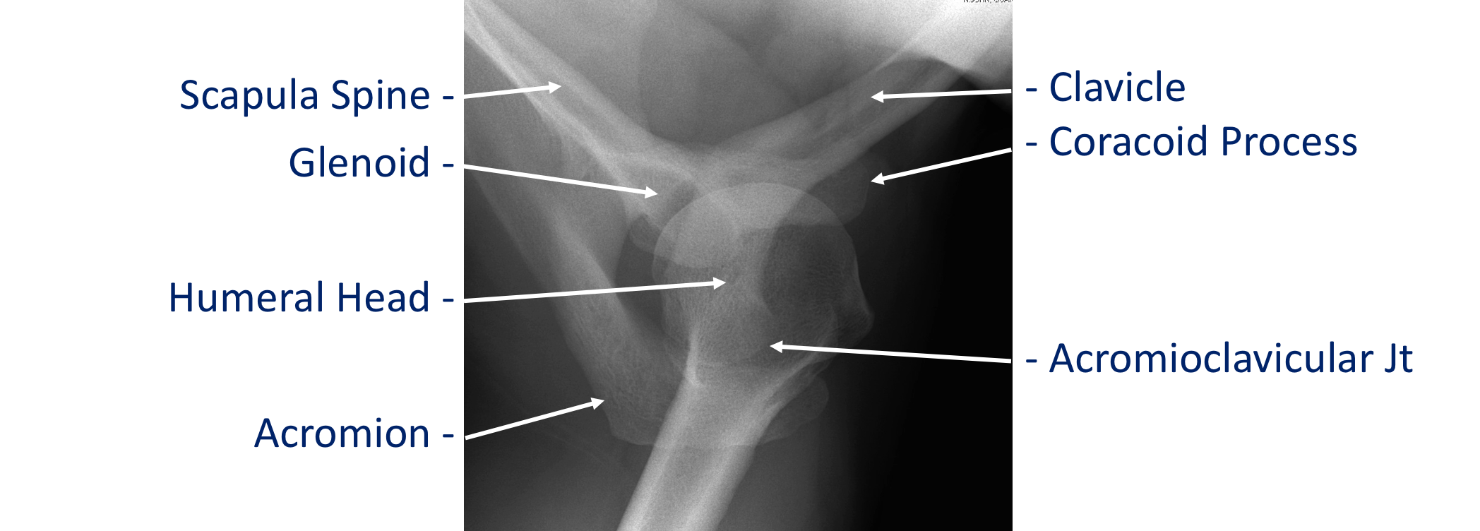
X-Rays are a quick and easy investigation to mainly look at the bones in the shoulder. They can demonstrate simple fractures and breaks and whether the joint is dislocated or not. They can pick up certain bone spurs and established arthritis. Whilst x-rays do not show soft tissue structures they can pick up areas of calcification which can occur in damaged ligaments and tendons.
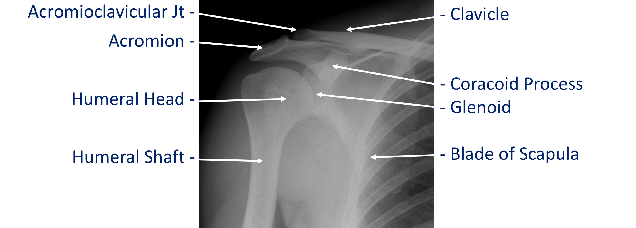
X-rays are usually taken from multiple angles to help look at the bones of the shoulder more clearly. However, due to its complex shape, x-rays of the shoulder may not be able to pick up all fractures. X-rays my also only give limited information in complex instability problems where there can be some loss of bone and in arthritis. In these situations, further investigations in the form of a CT or a MRI scan might be required.
X-rays do not show the soft-tissue structures around the shoulder. An MRI or Ultrasound scan is usually required to look at the muscles, ligaments or tendons in patients with rotator cuff or instability problems.

USS
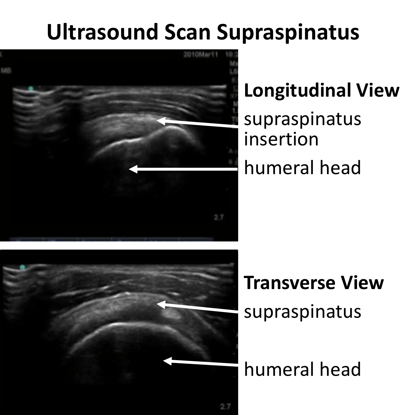
Ultrasound Scans for Shoulder Problems
Ultrasound scans use sound waves to produce images of the body. A transducer, which produces high frequency sound waves, is held against the skin. The sound waves penetrate the body and bounce back as echoes when they hit different structures. The returned sound waves are picked up by the transducer and can be processed to produce an image.
Ultrasound scans are particularly useful for looking at soft-tissues and the muscles, ligaments and tendons around the shoulder. They are also very good at looking at cysts, lumps and fluid filled collections.
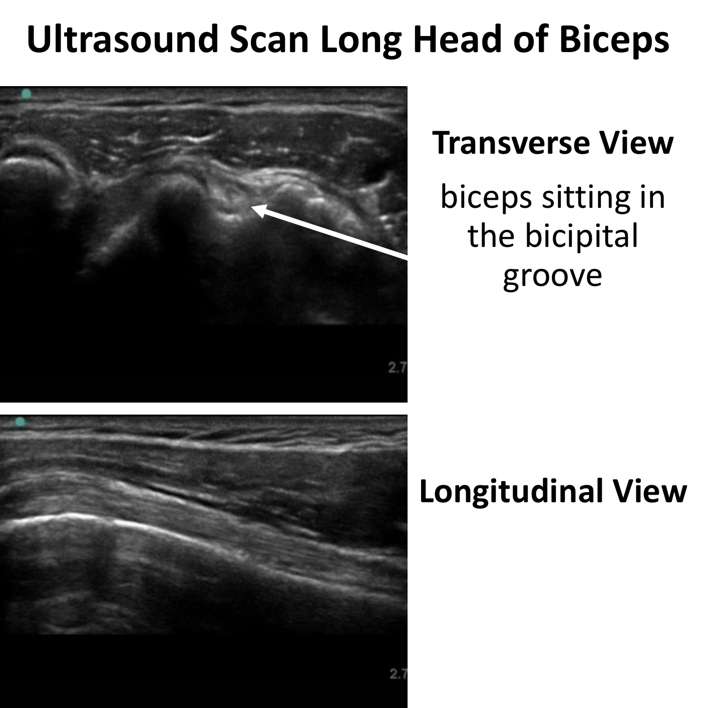
Ultrasound scans do not use any radiation and have no harmful effect. They have the added advantage of being able to look at ‘real-time’ / moving images. An ultrasound scan can look at the tissues as they are moving, which is particularly useful in the shoulder. In certain conditions they can demonstrate whether ligaments or tendons are moving over each other correctly or are being compressed as they move under a spur of bone.
A further benefit of being able to see moving structures is the ability to undertake ultrasound guided injections. In certain conditions, where pin-point accuracy is required, a needle can be directed by the physician into the exact area where the injection is required.
Ultrasound scans do not, generally, give very much information about bones.
MRI
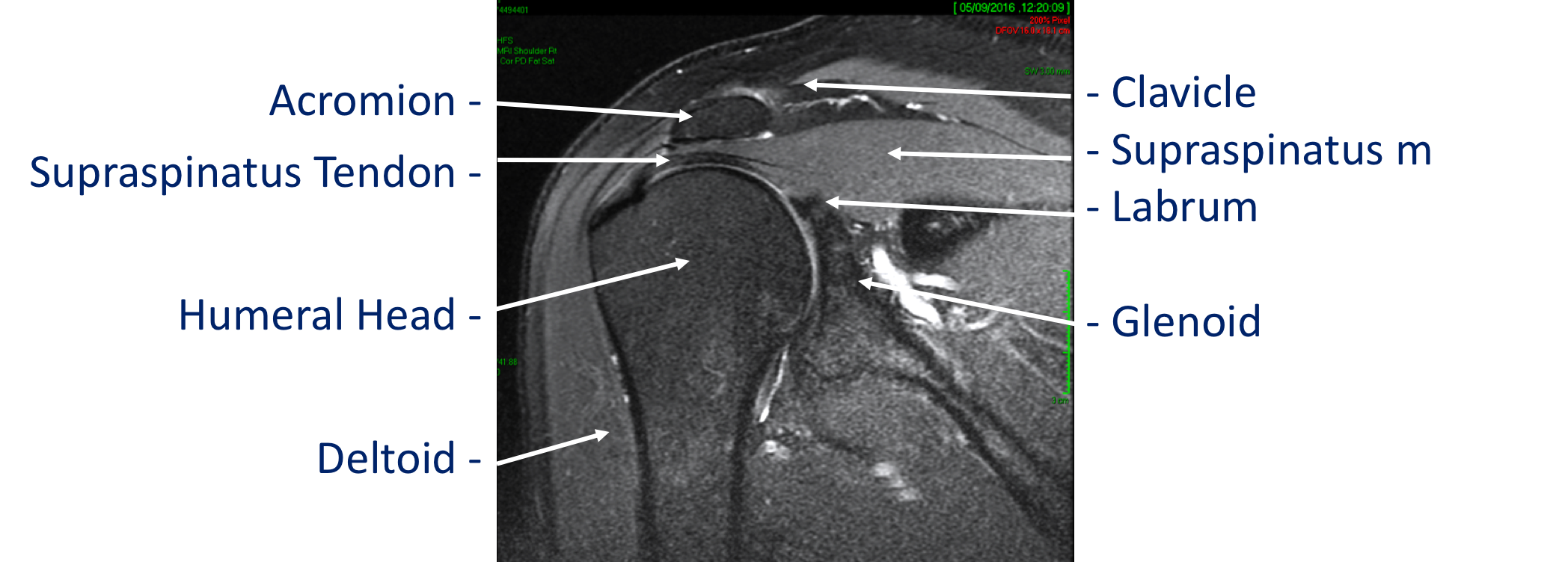
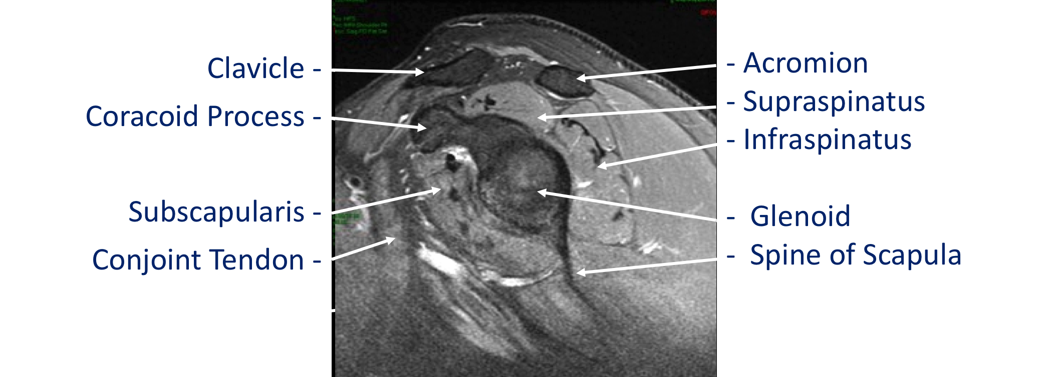
MRI Scan (Magnetic Resonance Imaging) for Shoulder Problems
MRI scanning is an imaging technique that is able to exploit the magnetic properties of hydrogen atoms within the body. It is able to produce very detailed images of multiple structures using magnetic fields and radio-waves without using any ionizing radiation.
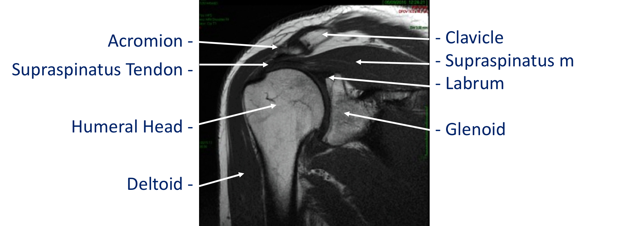
MRI scans do not use x-rays and have no harmful effects. They are particularly useful for looking at soft-tissue structures such as muscles ligaments and tendons and can also visualize articular cartilage. Whilst they do show boney structures plain x-rays and CT scans tend to be better for viewing bones.
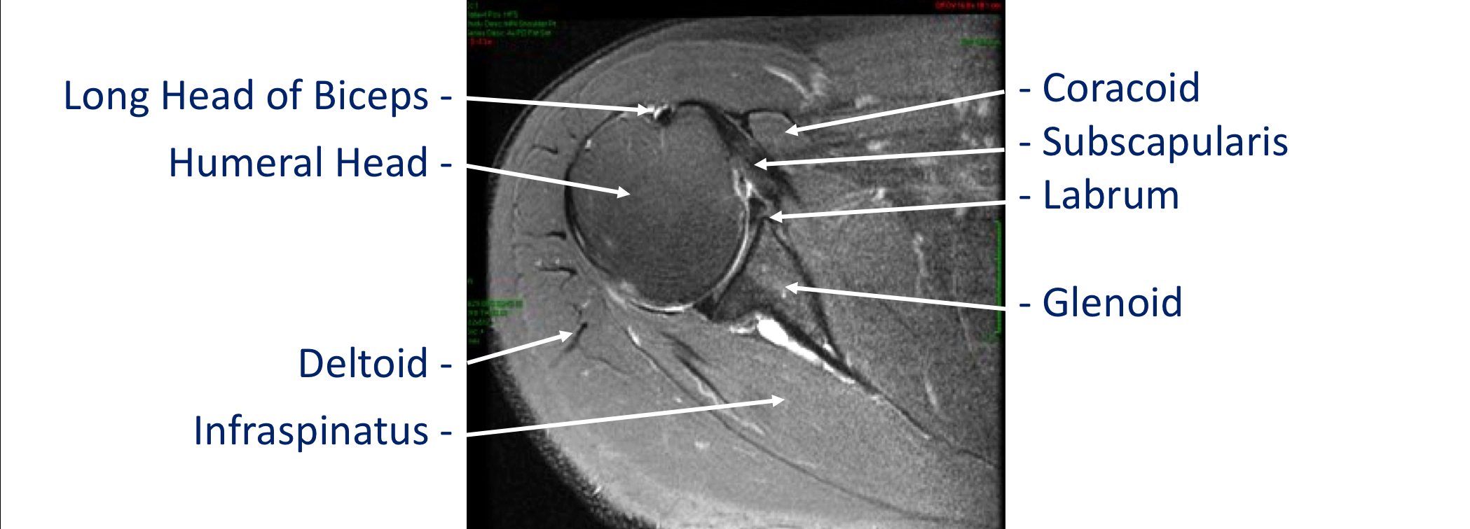
For shoulder problems MRI scans are particularly useful for looking at tendon and muscle damage in the rotator cuff and for ligament and labral injuries in instability. MRI scans are very good at looking at inflammation and in certain circumstances can give an indication as to how long an injury has been present.
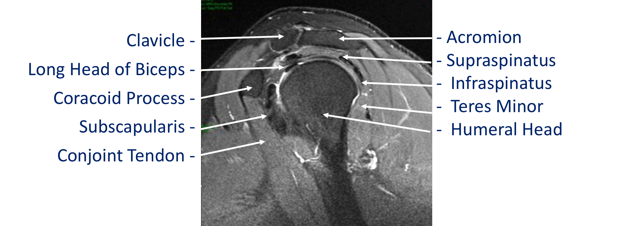
MRI scans can look at the shoulder from multiple angles and sometimes special views or sequences are used to look for particular problems. A special ‘dye’ called Gadolinium can help to highlight more clearly damage to the ligaments and labrum in patients with instability or SLAP tears. This can either be injected into the shoulder joint directly or through an intravenous injection in the arm.
CT Scans
CT Scan (Computed Tomography) for Shoulder Problems
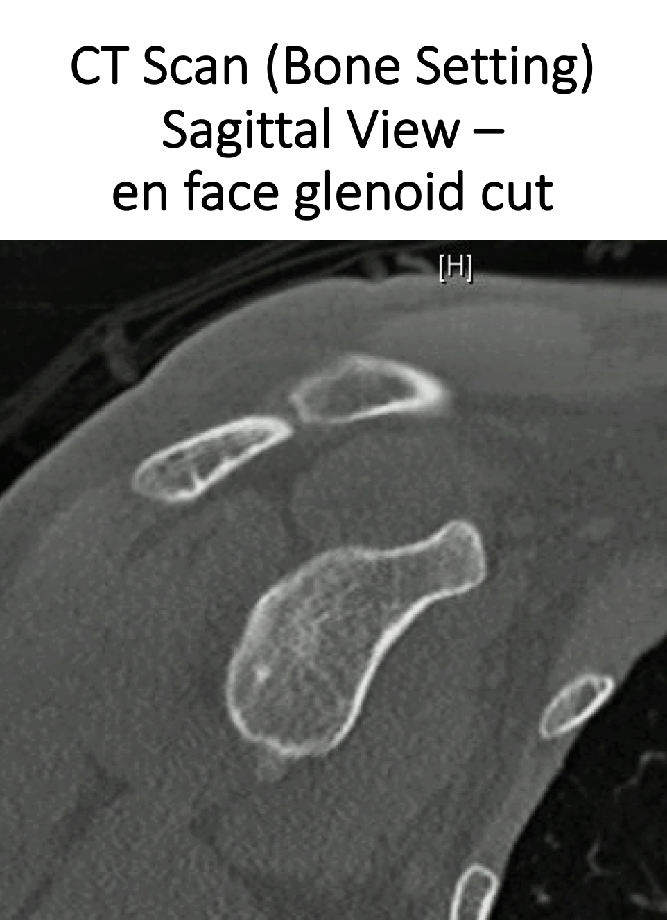
CT Scans are in fact multiple x-rays that are taken in rapid sequence as the scanner rotates around the patient. A computer is then able to process these and produce images in multiple plains and in 3-D. CT scans are very good at looking at bones which is particularly useful in the shoulder, where its complex shape can make it difficult to visualize using 2-D x-rays.
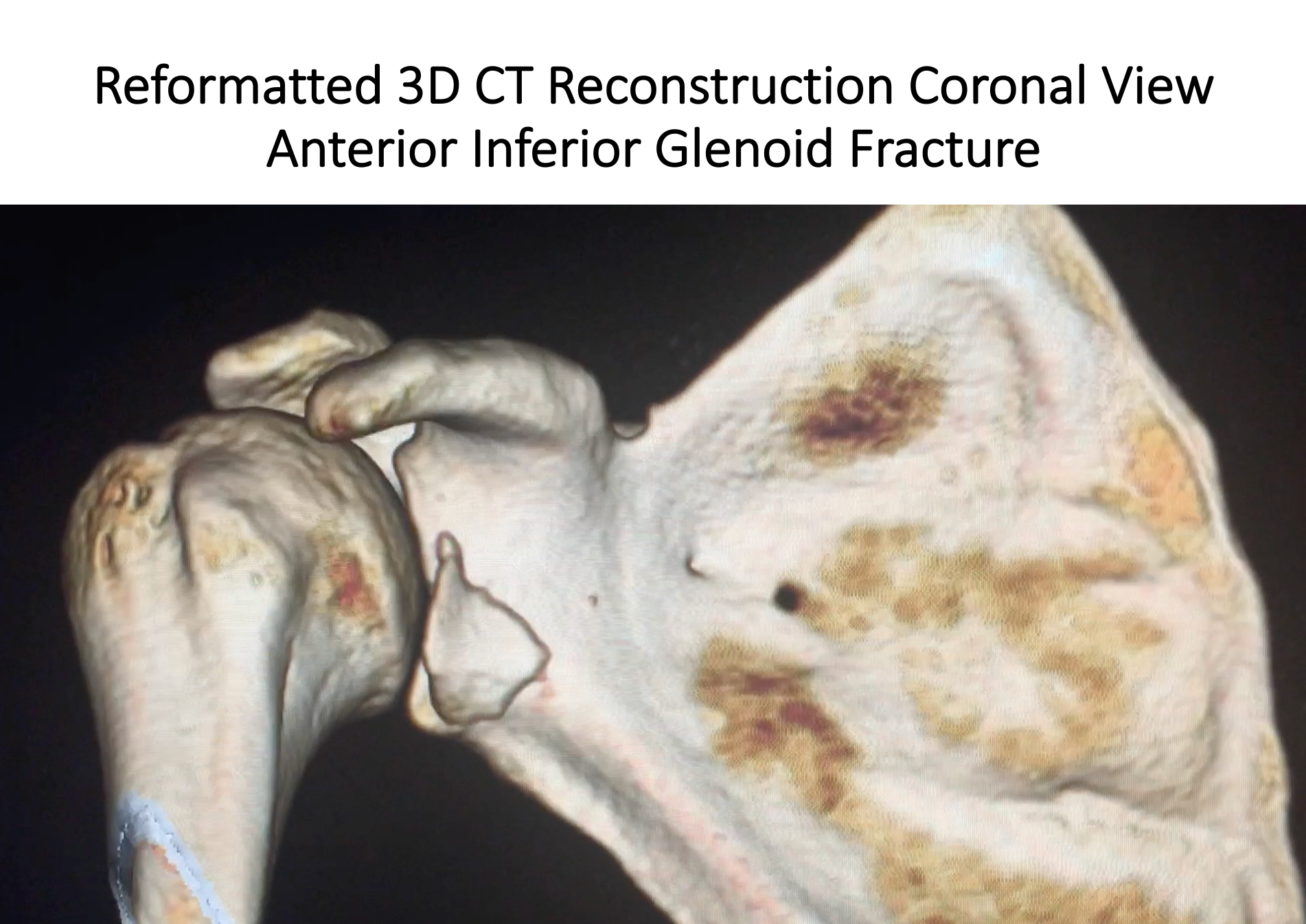
CT scans are used to look for fractures around the shoulder and can give valuable information as to the presence of a fracture, it’s size or shape. It can also indicate whether the fragments are in the correct position and whether a fracture is healing or has healed. The shape of the bones and any loss of bone can also be visualized with a CT scan. This can be useful in diagnosis and planning treatment with shoulder instability, arthritis or following injury.