Partial-Thickness Rotator Cuff Tears
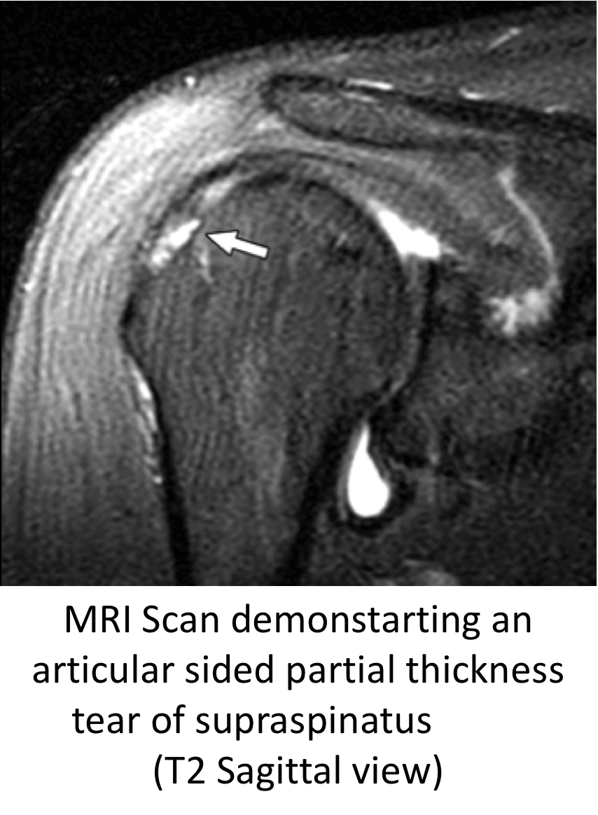
- Symptomatic Partial-Thickness Rotator Cuff Tears tend to occur in younger patients
- Unlike full-thickness tears they often occur in relatively healthy tendons
- They are often of some form of direct injury or an overuse injury, often related to sport
- Partial-Thickness tears can be particularly painful and maybe the result of the increased load on the remaining, untorn tendon fibres
Treatment of Partial Thickness Cuff Tears
- The initial treatment for a Partial-Thickness Rotator Cuff Tear, as for any rotator cuff tear is non-operative
- For patients who fail to respond to non-operative treatments or who symptoms recur, surgical repair is the best treatment
Arthroscopic Partial Thickness Rotator Cuff Repair
The best operation for someone with a symptomatic Partial Thickness Rotator Cuff Tear, that is refractory to treatment, is an Arthroscopic Partial Thickness Rotator Cuff Repair. This is an operation where the Arthroscopic camera is positioned and moved between the Glenohumeral Joint (Shoulder Joint) and the Subacromial Space above it. The Rotator Cuff Tendons can then be visualized from both sides and the damage identified. Using specialized instruments, the damaged tendon can be freshened and repaired back to its original bone origin, being held in place by specialized implants. During the repair a Subacromial Decompression is also undertaken to create more space for the repaired tendon to run underneath the Acromion.
find out more about Arthroscopic Shoulder Surgery and Implants…
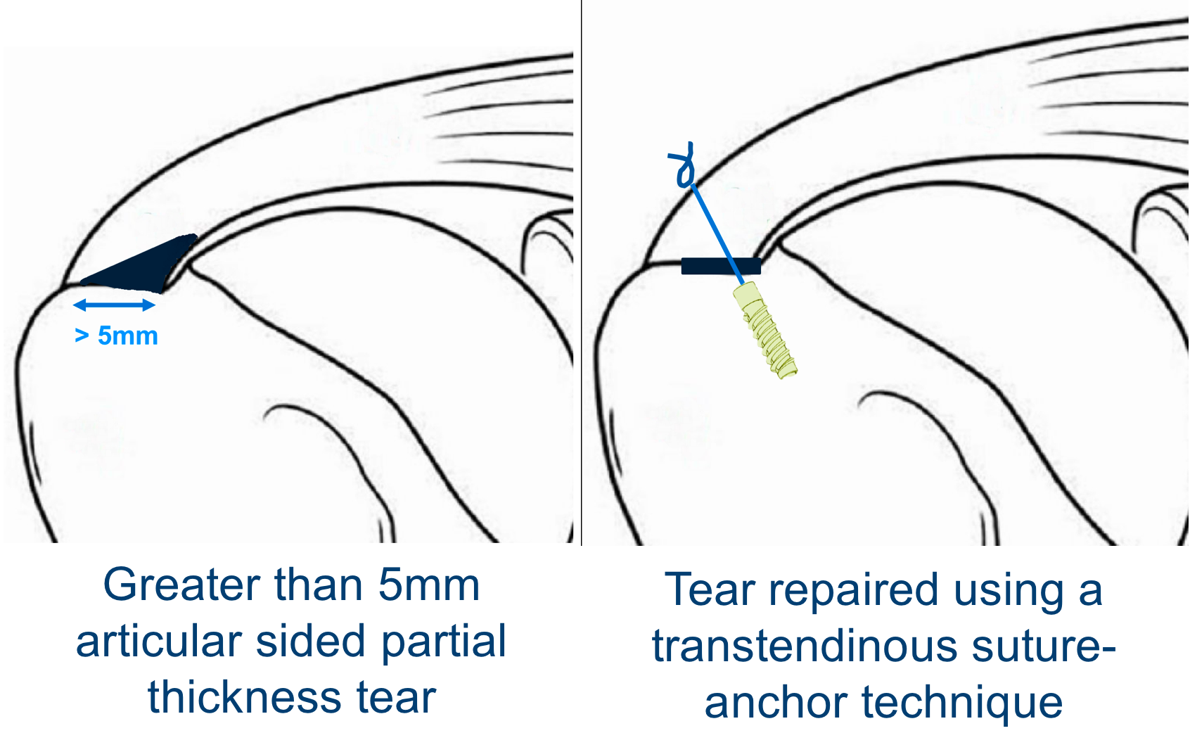
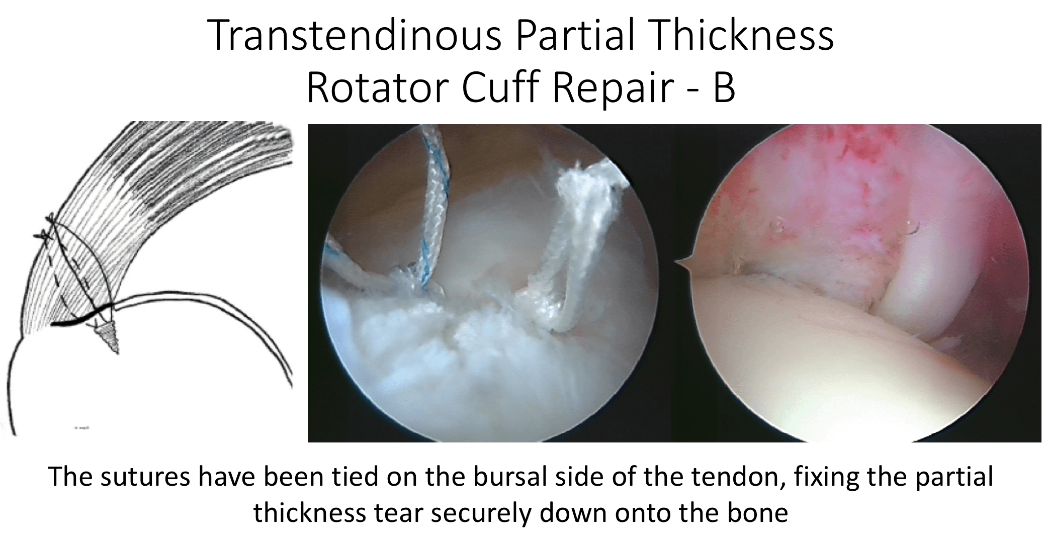
Depending on the exact position and size of the Partial Thickness Tear, the tendon can either be repaired using a transtendinous technique. If the tear as almost completely through the tendon it can be converted into a Full Thickness tear and then be repaired in that way. The specific implants that are used can also vary depending on certain technicalities and using implants that are the most appropriate. My routine Arthroscopic Trans-Tendinous Partial Thickness Rotator Cuff Repair Procedure is described below,
watch a video of a Partial Thickness Rotator Cuff Repair ….
- The patient is anaesthetised with a general anaesthetic and interscaelane nerve block
Find out more about having an anaesthetic….
Find out more about an Interscalene Nerve Block….
- A single prophylactic dose of broad-spectrum anti-biotics is administered
- A posterior, anterior and 1 – 3 lateral portals are used to access the Gleno-Humeral Joint (Shoulder Joint) and Subacromial spaces
- The Glenohumeral Joint is initially viewed to assess the exact position and size of the Rotator Cuff Tear and to look for any other associated problems, particularly with regards to the Long Head of Biceps
- The Subacromial Space is then entered and the superior surface of the Rotator Cuff and undersurface of the Acromion identified and cleared.
- With the arthroscope back in the Glenohumeral Joint a longitudinal split is made at the base of the Partial Tear
- The bone of the exposed Greater Tuberosity is then prepared and de-corticated.
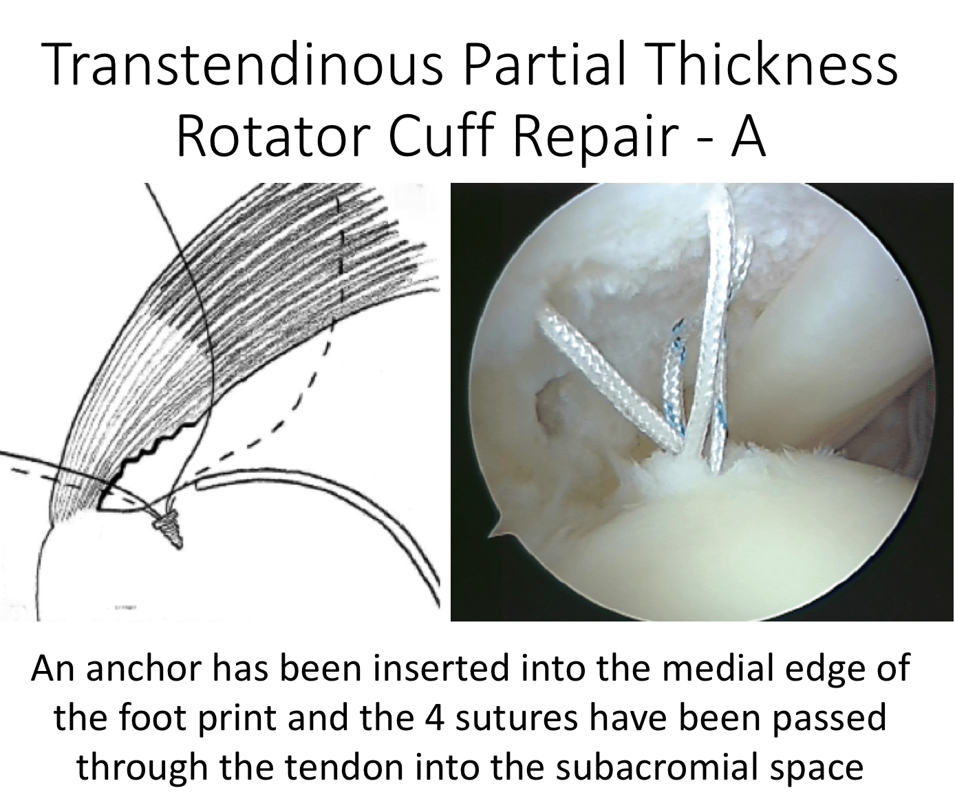
- An appropriate number of Full Thread, Bio-composite Anchors are then inserted into the medial edge of the prepared bone.
- The sutures from the anchors are then individually passed through the tendon from within the joint to the subacromial space
- With the arthroscope back in the Subacromial space the individual pairs of sutures are identified and tied down. This will anchor the torn part of the tendon back down onto the exposed bone
- At the end of the procedure a Subacromial Decompression is undertaken to create more clearance space for the repaired tendon to run underneath the Acromion.
- The Joint and the Subacromial Space is then washed out and the wounds closed with sub-cuticular sutures
After the Surgery
Post-Operative Care
Following an Arthroscopic Partial Thickness Rotator Cuff Repair the patient maybe able to go home on the same day as their surgery or may stay in the Hospital over-night. This would depend on various other factors. I would see the patient after the surgery to discuss how the procedure has gone and arrange for further Follow-Up. The patient will be seen by the In-Patient Physiotherapy team, who will instruct them on the initial Rehabilitation Protocol for their shoulder and how to use their Sling. Further Out-Patient physiotherapy will then be organised.
Find out more about Physiotherapy following Rotator Cuff Surgery….

I would usually review patients in the clinic 1 month and 3 months after their procedure to assess their progress and recovery.
Rehabilitation Protocol
Immediately after the surgery, when the patient has woken up from their general anaesthetic, their shoulder and arm will be numb from the interscalaene nerve block. This will usually last for 18 – 24 hours after the surgery. After the nerve block has worn off the arm can come out of the sling for washing and dressing and for certain controlled movements, as directed by the physiotherapists.
After a Rotator Cuff Repair, it is initially important to ‘protect’ the repair from any significant load as it begins to heal whilst, at the same time, it is preferable to not allow the shoulder to get too stiff. The physiotherapists will instruct the patient on the ‘safe zones’ of movement out of their sling over this period. Patients would usually need to have their arm in a sling for 2 weeks after a Partial Thickness Rotator Cuff Repair. Occasionally, this can vary depending on technical factors and tissue quality related to the surgery.
My standard rehabilitation protocol is outlined below. The information and time to recovery are a general estimation and may vary from person to person.
|
Post op |
|
| Immediate |
|
| Day 1-3
Weeks |
|
| 3-6 Weeks |
|
| 6 Weeks + |
|
| 12 Weeks |
|
|
Milestones |
|
|
Week 8 |
ROM 75%-80% of normal, sling completely discarded |
|
Week 12 |
Full ROM |
|
Week 20 |
Unrestricted activity |
|
Return to Functional Activities |
|
| Driving |
|
| Swimming |
|
| Golf |
|
| Heavy Lifting |
|
| Work |
|
| Manual - as guided by surgeon | |
Success of Surgery, Risks & Complications
An Arthroscopic Partial Thickness Rotator Cuff Repair is usually a very successful procedure with regards to alleviating pain and restoring good function. The surgery is aimed at freshening up the damaged tendon and its bone insertion and then fixing it back into position under no tension. At this point the ‘degenerate’ tendon can begin to try and heal. After this it can take more than 4 months for the healing process to be completed and, even at the end of that, the healed tendon will not be as good as new. As a result, it can take 6 months or more for the shoulder to be as good as it is going to be and the repaired Rotator Cuff will never be as strong as a 21 year olds. There is a danger of the Tendon failing to heal or re-tearing after a Partial Thickness Rotator Cuff Repair. However, this strangely does not always mean that the symptoms will return!
At 6 months after a Partial Thickness Rotator Cuff Repair, about 75-80% of patients have an excellent result, 10-15% of patients still have occasional problems with their shoulder, but are pleased that they have had surgery and about 5% of patients do not feel that their shoulder has significantly improved, although it is no worse than before.
There are always risks and complications associated with any operation.
- Anaesthetic - The risks of having a General Anaesthetic and an Interscalaene Nerve Block are very low, but will always need to be assessed on an individual basis by an Anaesthetist. Suffice it to say, that whilst a Shoulder Operation can in no way be considered a ‘life-saving’ procedure, an Anaesthetist would not consider undertaking an anaesthetic if they had any concerns that an undue risk was being taken
- Re-Tear / Failure to Heal – About 20% of tendons fail to heal after a Rotator Cuff Repair. However they are NOT always symptomatic
- Infection – Infection following arthroscopic surgery is rare < 0.2%
- Neurovascular Injury – Damage tosignificant neurovascular structures during arthroscopic shoulder surgery is rare < 0.2%
- CRPS Type 1 – A Chronic Pain Syndrome following arthroscopic shoulder surgery is rare < 0.2%
Outcome Measures
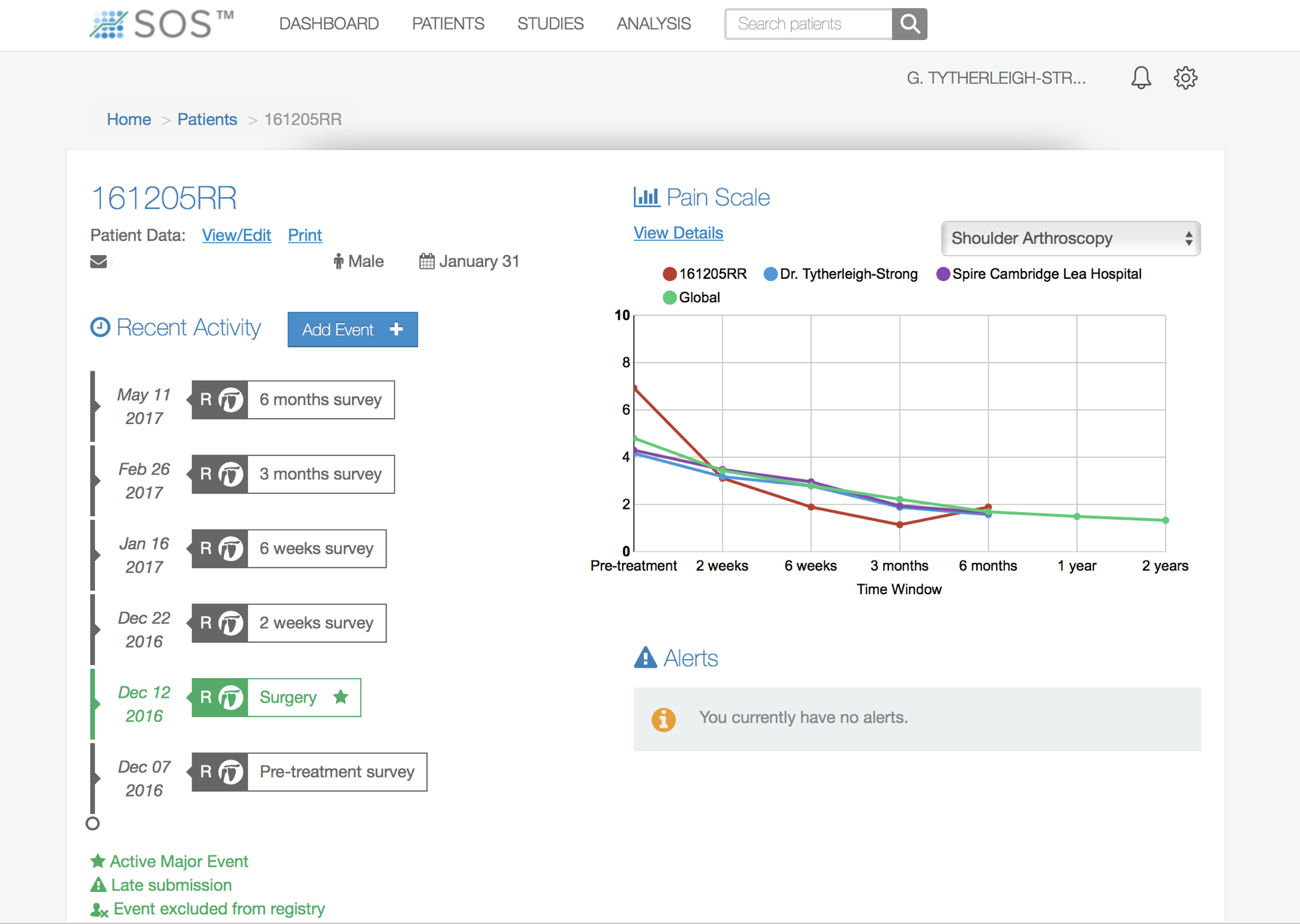
Assessing patient outcomes following surgery, using validated scoring systems, is a very important and useful exercise.
find out more about outcome measures and the SOS system….(Patient Information – Outcome Measures)
Partial-Thickness Rotator Cuff Tears
-
There is a vast variation between the specific symptoms, types and chronicity of a tear and the needs and expectations of patients who present with a symptomatic Rotator Cuff Tear
-
There are also many different ways of treating and repairing Rotator Cuff Tears
-
My approach to treating a patient with a symptomatic Rotator Cuff Tear is to assess the specific issues and nature of their problem and to then base my recommendation of how to treat their Rotator Cuff Tear, based on this
Surgery for Rotator Cuff Tears
-
I undertake all of my Rotator Cuff Repairs using Arthroscopic Surgery (keyhole surgery)
-
The basic aim of any Rotator Cuff Repair is to mobilise and freshen up the ends of the torn tendon, to freshen up the boney insertion on the humerus and then to position and re-attach the tendon back down to its original insertion, achieving a tensionless repair
-
To achieve this there are multiple techniques, implants and strategies that can be used
-
The descriptions of the procedures outlined below are based on the general technique. The specific details and pros and cons of the various implants and equipment that I use are covered in the Arthroscopic Surgery Section
Healing of Rotator Cuff Tears
-
Rotator Cuff Tears usually occur in tendons that have undergone ‘wear and tear’ and, as the tear occurs, the degenerate tendon is no longer strong enough to withstand the mechanical forces placed on it
-
When a Rotator Cuff Tendon is repaired it is still degenerate and, as a result, its healing potential is not as good as a normal, healthy tendon
-
Surgical techniques and implant technology are constantly advancing and we are currently able to mobilise and successfully repair nearly all tendon tears
-
However, despite this, a number of repairs still fail to heal
-
This is likely to be due to the poor healing potential of degenerate tendons
-
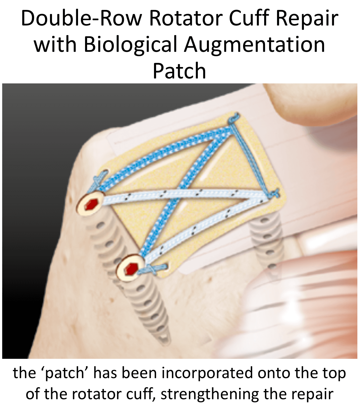
The latest advance in Rotator Cuff Repairs, ‘Ortho-Biologics’, involves trying to improve and optimize the healing potential of degenerate tendons
-
Although ‘Ortho-Biologics’ is in its infancy, it is an area where I have a great interest and I incorporate its use, where indicated, during Rotator Cuff surgery
find out more about ‘Ortho-Biologics’…..
Full Thickness Rotator Cuff Repair
The best operation for someone with a symptomatic repairable Full Thickness Rotator Cuff Tear, that is refractory to treatment, is an Arthroscopic Rotator Cuff Repair. Full Thickness Rotator Cuff Tears can be acute or chronic, come in all shapes and sizes and can occur in tendons that have undergone varying amounts of degeneration. As a result, every Rotator Cuff Tear is slightly different.
In response to this, the exact technique, type and number of implants and potential beneficial use of ‘Ortho-Biologics’ is something that I consider individually for each Rotator Cuff tear before undertaking a surgical repair. Described below is the general operative technique that I use to undertake an Arthroscopic Rotator Cuff Repair, although this often varies slightly depending on the exact nature of an individual tear,
Arthroscopic Rotator Cuff Repair
watch a video of a Rotator Cuff Repair …
Find out more about arthroscopic surgery….
- The patient is anaesthetised with a general anaesthetic and interscaelane nerve block
- A single prophylactic dose of broad-spectrum anti-biotics is administered
- A posterior, 1 – 2 anterior and 1 – 2 lateral portals are used to access the Gleno-Humeral Joint (Shoulder Joint) and Subacromial spaces
- The Glenohumeral Joint is initially viewed to assess the exact position and size of the Rotator Cuff Tear and to look for any other associated problems, particularly with regards to the Long Head of Biceps
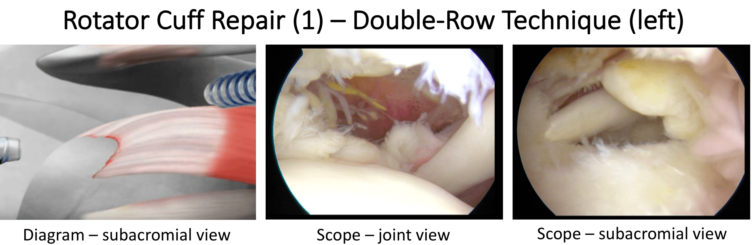
- The Subacromial Space is then entered and the Rotator Cuff Tear identified and assessed for its size, shape and tissue quality
- Usually, at the outset of the procedure, a Subacromial Decompression is performed to create more clearance space to undertake the repair and then to protect the repair, after completion
- The torn tendon is then fully mobilised and it edges freshened
- The exposed bony Foot-Print of the tendon insertion on the Greater Tuberosity is then prepared and de-corticated down to bleeding cancellous bone
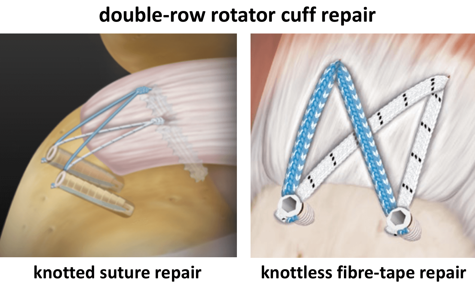
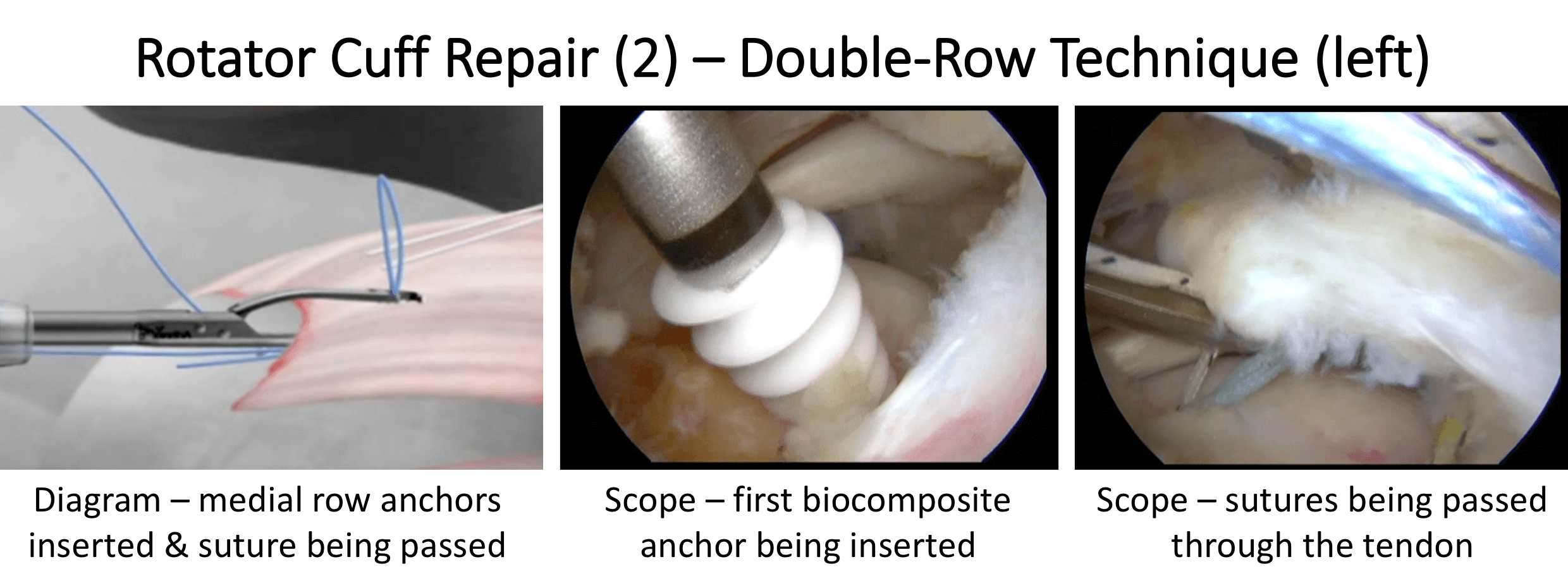
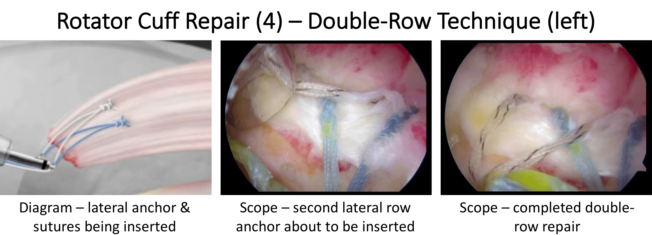
- A combination of Full Thread, Bio-Composite Anchors are then inserted, usually in a double-row configuration, as is considered appropriate to obtain the best repair.
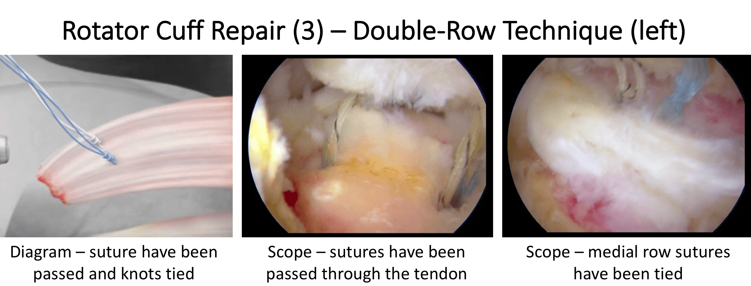
- The sutures from the anchors are then individually passed through the tendon, tied and secured to anchor it securely down to the bone
- Where considered appropriate, some form of ‘Ortho-Biologics’ may also be used to help promote tendon healing
- At the end of the procedure the Repair is assessed, the joint and subacromial space washed out and the wounds closed with sub-cuticular sutures
After the Surgery
Post-Operative Care
Following an Arthroscopic Rotator Cuff Repair the patient maybe able to go home on the same day as their surgery or may stay in the Hospital over-night. This would depend on various other factors. I would see the patient after the surgery to discuss how the procedure has gone and arrange for further Follow-Up. The patient will be seen by the In-Patient Physiotherapy team, who will instruct them on the initial Rehabilitation Protocol for their shoulder and how to use their Sling. Further Out-Patient physiotherapy will then be organised.
Find out more about Physiotherapy following Rotator Cuff Surgery….
I would usually review patients in the clinic 1 month and 3 months after their procedure to assess their progress and recovery.
Rehabilitation Protocol
Immediately after the surgery, when the patient has woken up from their general anaesthetic, their shoulder and arm will be numb from the interscalaene nerve block. This will usually last for 18 – 24 hours after the surgery. After the nerve block has worn off the arm can come out of the sling for washing and dressing and for certain controlled movements, as directed by the physiotherapists.
After a Rotator Cuff Repair, it is initially important to ‘protect’ the repair from any significant load as it begins to heal whilst, at the same time, it is preferable to not allow the shoulder to get too stiff. The physiotherapists will instruct the patient on the ‘safe zones’ of movement out of their sling over this period. Patients would usually need to have their arm in a sling for 2 weeks after a Partial Thickness Rotator Cuff Repair. Occasionally, this can vary depending on technical factors and tissue quality related to the surgery.
My standard rehabilitation protocol is outlined below. The information and time to recovery are a general estimation and may vary from person to person.
|
Post op |
|
| Immediate |
|
| Day 1-3
Weeks |
|
| 3-6 Weeks |
|
| 6 Weeks + |
|
| 12 Weeks |
|
|
Milestones |
|
|
Week 8 |
ROM 75%-80% of normal, sling completely discarded |
|
Week 12 |
Full ROM |
|
Week 20 |
Unrestricted activity |
|
Return to Functional Activities |
|
| Driving |
|
| Swimming |
|
| Golf |
|
| Heavy Lifting |
|
| Work |
|
| Manual - as guided by surgeon | |
Success of Surgery, Risks & Complications
An Arthroscopic Rotator Cuff Repair is usually a very successful procedure with regards to alleviating pain and restoring good function. The surgery is aimed at freshening up the damaged tendon and its bone insertion and then fixing it back into position under no tension. At this point the ‘degenerate’ tendon can begin to try and heal. After this it can take more than 4 months for the healing process to be completed and, even at the end of that, the healed tendon will not be as good as new. As a result, it can take 6 months or more for the shoulder to be as good as it is going to be and the repaired Rotator Cuff will never be as strong as a 21 year olds. There is a danger of the Tendon failing to heal or re-tearing after a Rotator Cuff Repair. However, this strangely does not always mean that the symptoms will return!
At 6 months after a Rotator Cuff Repair, about 75-80% of patients have an excellent result, 10-15% of patients still have occasional problems with their shoulder, but are pleased that they have had surgery and about 5% of patients do not feel that their shoulder has significantly improved, although it is no worse than before.
There are always risks and complications associated with any operation.
-
Anaesthetic - The risks of having a General Anaesthetic and an Interscalaene Nerve Block are very low, but will always need to be assessed on an individual basis by an Anaesthetist. Suffice it to say, that whilst a Shoulder Operation can in no way be considered a ‘life-saving’ procedure, an Anaesthetist would not consider undertaking an anaesthetic if they had any concerns that an undue risk was being taken
-
Re-Tear / Failure to Heal – About 20% of tendons fail to heal after a Rotator Cuff Repair. However they are NOT always symptomatic
-
Infection – Infection following arthroscopic surgery is rare < 0.2%
-
Neurovascular Injury – Damage to significant neurovascular structures during arthroscopic shoulder surgery is rare < 0.2%
-
CRPS Type 1 – A Chronic Pain Syndrome following arthroscopic shoulder surgery is rare < 0.2%
Outcome Measures
Assessing patient outcomes following surgery, using validated scoring systems, is a very important and useful exercise.
Irreparable Rotator Cuff Tears Non-Operative Management
Irreparable Rotator Cuff Tears Non-Operative Management:
Deltoid Re-Training Programme
- A Deltoid Re-Training Programme is aimed at strengthening and re-training the Deltoid and other muscles around the shoulder to try and compensate for the absent Rotator Cuff muscles
- It works by re-training these muscles to help re-centralise and hold the Humeral Head into the Glenoid Socket and allow the other muscles around the shoulder to work properly
- The Deltoid Re-Training Programme is a set of relatively simple and easy exercises that need to be repeated every day
- However, it can take many months to gain a good result. This is something that would initially best be supervised by a Physiotherapist
- Sometimes pain and discomfort can be a big problem and significantly hamper a patient’s ability to do their exercises
- If this is the case a Subacromial Cortisone injection or a temporary Suprascapular nerve block can help
- Patients can often obtain quite a good function and range of motion following Deltoid Re-Training
- However, their strength is often not particularly good and their shoulder muscles often fatigue quite quickly
Pain Management
Analgesia/Pain Relief
- pain is the most severe problem associated with a Frozen Shoulder, particularly in the Freezing Stage
- there are no medals for bravery for a patient with a Frozen Shoulder
- NSAID (Non-Steroidal Anti-Inflammatories) –
- NSAIDs work be reducing the painful inflammatory response, they are useful at all Stages of Frozen Shoulder
- NSAIDs can damage the stomach lining and affect the kidneys. It is important that a patient’s Family Doctor prescribes this medication if it is going to be used for a longer term
- Codeine based Analgesics –
- Codeine based analgesics are pain killers and affect a patient’s perception of pain. As a result, they can have some effect on consciousness depending on their strength
- Codeine based analgesics can lead to constipation if taken for a longer time. Having a high-fibre diet or even taking laxatives might need to be considered
- Nociceptive Analgesics –
- Nociceptive pain killers work on nerve generated pain
- Amitriptyline in lower doses works as a nociceptive pain killer. It has a useful side-effect in that it can make patients drowsy
- in cases of severe pain Amitriptyline can be prescribed at night
Subacromial Cortisone Injection –
- If someone’s Supraspinatus Tendonitis has failed to settle, after an adequate course of Physiotherapy, a Subacromial Cortisone injection can help to settle the symptoms down. Cortisone is a corticosteroid that is naturally produced by the body’s Adrenal Gland. Injectable Cortisone is synthetically produced and has a very powerful ant-inflammatory action. When injected into the Subacromial space it has the potential of settling severe inflammation, allowing the patient to undertake their rehabilitation exercises.
- A Subacromial Cortisone can be easily administered in the Out-Patient Clinic. It is a quick and relatively painless procedure. Afterwards patients can continue with their normal day-to-day activities. Occasionally patients can feel a bit of soreness around their shoulder later that day, but this usually passes fairly quickly.
- It often takes several days before someone notices the benefits following a Cortisone injection and sometimes several weeks. The Cortisone works in the background and there is no specific requirement to particularly rest the shoulder or to do extra exercises. The full benefits of a Cortisone injection are usually felt within a month. In some cases, the Cortisone may not give any benefit, this may be an indication of the severity of the Supraspinatus Tendonitis.
- A Cortisone injection only lasts in the body for a few days. Any benefit that someone gets from the Cortisone will be from its acute anti-inflammatory effect allowing the Supraspinatus Tendonitis to settle. If the symptoms do return after a while, it is not because the Cortisone has worn off, but because the inflammation has returned.
- Infections can occur after any type of injection but are extremely rare (1 in 15,000). If someone feels that they are developing an infection within 48 hours of a Cortisone injection they should seek advice from their Family Practitioner.

Suprascapular Nerve Block
- Occasionally a patient can have a shoulder problem that cannot be treated by surgery or continues to cause severe pain despite the mechanical problem having been adequately treated.
- In this situation, it is possible to ‘block’ the nerve that supplies this area from registering pain, whilst the actual problem remains untreated.
- When a nerve is ‘blocked’ it stops working completely, so this option is only possible for certain nerves that supply a very specific area
- The commonest nerve block around the Shoulder is a Suprascapular Nerve Block
- Suprascapular Nerve Block
- The Suprascapular Nerve supplies the motor power for the supraspinatus and infraspinatus muscles and most of the pain sensation for the shoulder
- A selective Suprascapular Nerve Block can alleviate a lot of the pain sensation from around the shoulder but does have some effect on muscle function
- However, in patients with massive Rotator Cuff Tears and with severe arthritis the loss of muscle function has negligible effect
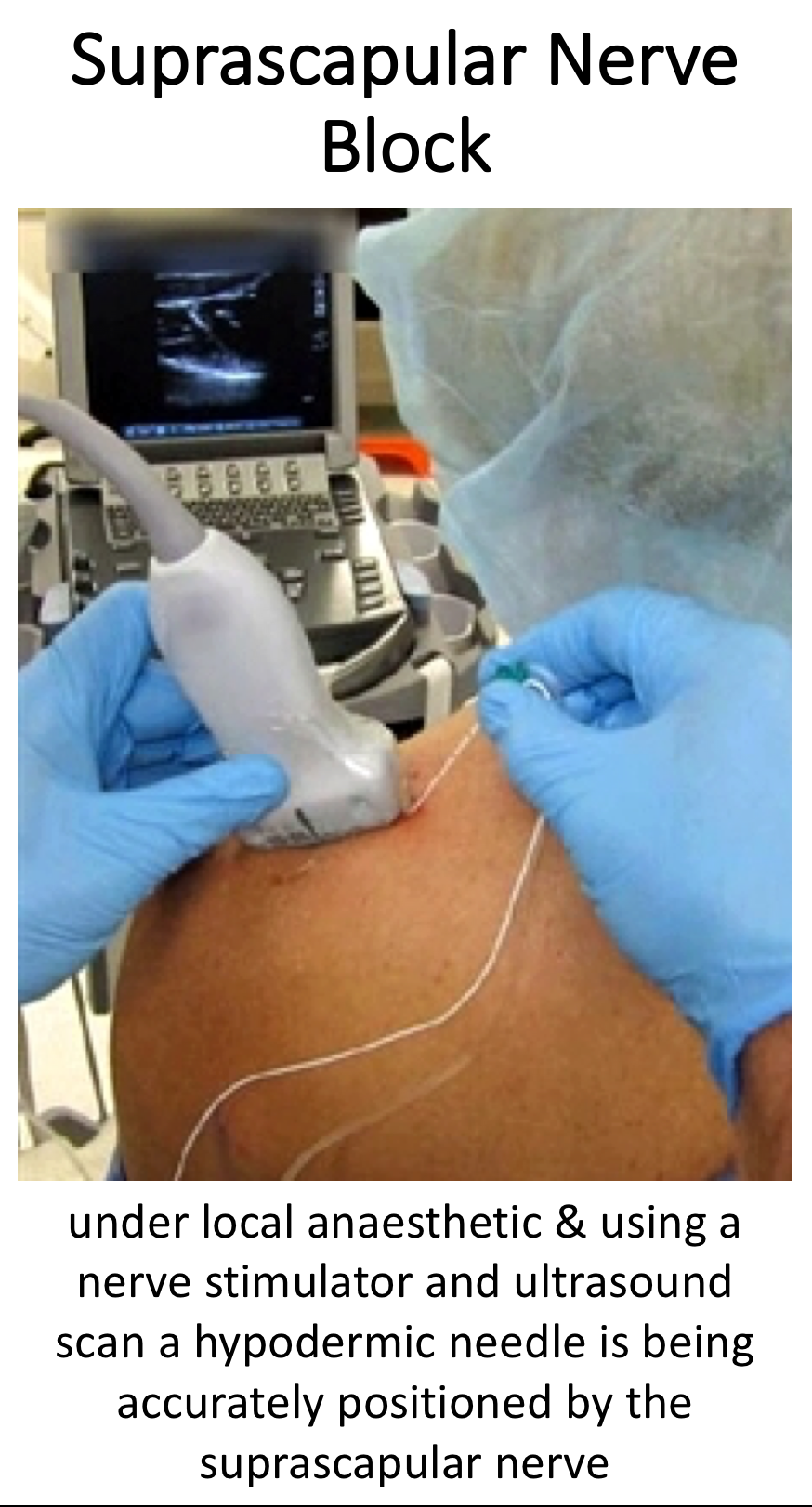
- A Suprascapular Nerve Block is undertaken by an Aneasthetist who specializes in nerve blocks
- The procedure is usually done as a Day Case procedure
- An Ultrasound Scan and nerve stimulator are used to accurately locate and direct a hypodermic needle to the Suprascapular Nerve where it passes through the Suprascapular Notch
- Long acting local anaesthetic is then injected around the nerve
- An initial nerve block is always temporary and will last about 3 months, this is to make sure that there are no unwanted ‘side-effects’
- A more permanent ‘nerve block’ can then be undertaken
Surgery for Irreparable Rotator Cuff Tears
Surgery for Irreparable Rotator Cuff Tears
For patients who would like to try and achieve as good a function as possible and for those who have not been helped by Deltoid Re-Training, there are a number of different operative procedures that are available to treat Irreparable Rotator Cuff Tears. These can range from simple arthroscopic procedures, more complex arthroscopic reconstructions, large tendon transfers to specialised shoulder replacements. Each procedure involves varying levels of difficulty, varying levels of success, varying levels of functional recovery and varying levels of risks and complications. I consider it very important that all of these factors are taken into consideration, along with a realistic expectation of success, when discussing with a patient which procedure is likely to give them the best result.
- There are a number of different procedures available for patients with symptomatic Irreparable Rotator Cuff tears.
- They vary in complexity and in the functional recovery that they are likely to produce.
- However, none of them are able to fully return a patient’s shoulder completely back to normal.
Arthroscopic Debridement (for Irreparable Rotator Cuff Tears)
An Arthroscopic Debridement is aimed at tidying up and removing all of the debris and damaged tissue from around the shoulder. It is primarily aimed at pain relief but often, as a result of reducing the level of pain, the patient’s shoulder function can be improved. In some cases, the Long Head of Biceps tendon, where it is still present, can be an additional source of pain and a Tenotomy (release of the tendon) may be of benefit. In other instances a component of someone’s pain may be due to irritation of the Suprascapular Nerve, addressing this with a Nerve Release or Ablation may also be of benefit. Both of these procedures can be done at the same time as an Arthroscopic Debridement.
An Arthroscopic Debridement is a relatively small procedure which can often be done as a Day-Case. The shoulder does NOT need to be immobilised after the surgery and patients can often notice an improvement fairly quickly.
However, like most operations for Irreparable Rotator Cuff Tears, results can be variable and it is not usually possible to predict how much any one person is likely to benefit from surgery until it has be done. My routine Arthroscopic Trans-Tendinous Partial Thickness Rotator Cuff Repair Procedure is described below,
find out more about the Suprascapular nerve….
Find out more about Arthroscopic Surgery….
Watch a video of an arthroscopic debridement of the shoulder….
Arthroscopic Debridement (+/- Biceps Tenotomy & Suprascapular Nerve Ablation)
- The patient is anaesthetised with a general anaesthetic and interscaelane nerve block
Find out more about having an anaesthetic….
Find out more about an Interscalene Nerve Block….
- A posterior, anterior and 1 – 2 lateral portals are used to access the Gleno-Humeral Joint (Shoulder Joint) and Subacromial spaces
- The Glenohumeral Joint is initially viewed to assess the size and extent of the tear and to look for any other associated problems, particularly with regards to Osteoarthritic changes to the joint and problems with the Long Head of Biceps
- Using a Shaver and Radiofrequency wand the joint is then cleaned out removing any loose fragments and excising any ragged tissue
- If the Long Head of Biceps is considered to be damaged a Tenotomy is performed
- The Subacromial Space is then entered and, using the Shaver and Wand, the stump of the torn Rotator Cuff Tendons are tidied up and any other ragged tissues excised
- If the Suprascapular is considered to be a source of pain it is exposed using an accessory Nervaiser type portal. The nerve is then either released or ablated
- At the end of the procedure the joint and subacromial space are assessed, washed out and the wounds closed with sub-cuticular sutures
After the Surgery
Post-Operative Care
Following an Arthroscopic Debridement the patient maybe able to go home on the same day as their surgery or may stay in the Hospital over-night. This would depend on various other factors. I would see the patient after the surgery to discuss how the procedure has gone and arrange for further Follow-Up. The patient will be seen by the In-Patient Physiotherapy team, who will instruct them on the initial Rehabilitation Protocol for their shoulder and will organise for further Out-Patient physiotherapy.
Find out more about Physiotherapy following Rotator Cuff Surgery….
I would usually review patients in the clinic 1 month and 3 months after their procedure to assess their progress and recovery.
Rehabilitation Protocol
Immediately after the surgery, when the patient has woken up from their general anaesthetic, their shoulder and arm will be numb from the interscalaene nerve block. This will usually last for 18 – 24 hours after the surgery. The patient’s arm will be in a Sling for this period to protect their arm. After the nerve block has worn off the arm can come out of the sling and can be used as normal.
After an Arthroscopic Debridement patients are encouraged to try and use and move their arm as much as possible, within the limits of discomfort. No specific surgical repairs have been undertaking, so there is no danger of damaging anything in the shoulder in using it. We advise patients to keep their Sling so that it can be occasionally used if their shoulder gets at all ‘tired’. We also recommend that patients consider using the Sling for ‘Social Protection’. Sometimes when people are out in public, wearing a Sling can warn people that a patient has had recent surgery and prevent them from inadvertently knocking that arm.
My standard rehabilitation protocol is outlined below. The information and time to recovery are a general estimation and may vary from person to person.
|
Post op |
|
| Immediate |
|
| Day 1-3
Weeks |
|
| 3-6 Weeks |
|
|
Return to Functional Activities |
|
| Driving |
|
| Swimming |
|
| Golf |
|
| Racquet Sports/Repeated |
|
| Overhead Activities | |
| Lifting | As able |
| Work | Sedentary - As able |
| Manual - 6 weeks, may need to modify activity for 3 months | |
Success of Surgery, Risks & Complications
An Arthroscopic Debridement is a safe and is usually a very successful procedure with regards to alleviating pain and restoring variable function. Whilst some people notice a significant benefit within weeks of surgery it can take many months for others. It is extremely rare that anyone’s shoulder is ever any worse than before the surgery.
There are always risks and complications associated with any operation.
- Anaesthetic - The risks of having a General Anaesthetic and an Interscalaene Nerve Block are very low, but will always need to be assessed on an individual basis by an Anaesthetist. Suffice it to say, that whilst a Shoulder Operation can in no way be considered a ‘life-saving’ procedure, an Anaesthetist would not consider undertaking an anaesthetic if they had any concerns that an undue risk was being taken
- Infection – Infection following arthroscopic surgery is rare < 0.2%
- Neurovascular Injury – Damage tosignificant neurovascular structures during arthroscopic shoulder surgery is rare < 0.2%
- CRPS Type 1 – A Chronic Pain Syndrome following arthroscopic shoulder surgery is rare < 0.2%
Outcome Measures
Assessing patient outcomes following surgery, using validated scoring systems, is a very important and useful exercise.
Superior Capsular Reconstruction
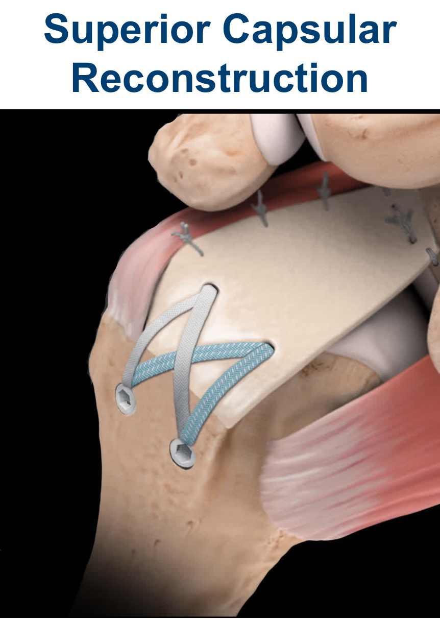
This is a very exciting new procedure that uses a Human Allograft Patch to reconstruct the Superior Capsule in ‘certain’ patients that have an Irreparable Rotator Cuff Tear. Using a fairly complex Arthroscopic technique, a very strong 4mm thick Patch is fixed between the edge of the Glenoid Socket and the Humeral Head using a combination of Anchors to secure the patch. It effectively reconstructs the Superior Capsule of the Shoulder and appears to re-centralise the Humeral Head and allow the other muscle groups around the shoulder to work effectively.
However, not every Irreparable Rotator Cuff Tear is suitable for this procedure. If there is complete or significant damage to the Infraspinatus Tendon or if the Subscapularis Tendon is significantly damaged a Superior Capsule Reconstruction is very unlikely to work. In addition, it is a big operation with a protracted recovery and may not be the best option for every patient.
Previously there were really no reliably successful surgical procedures available to improve ‘function’ for patients with a symptomatic Irreparable Rotator Cuff Tear. Although it is relatively early days, a Superior Capsule Reconstruction appears to be able to achieve this, certainly initially, when performed correctly on appropriate patients. I, personally, have been very pleased and impressed with the initial outcomes using this procedure on a limited number of patients. However, it is a new procedure and, as of yet, we do not know what will happen with patients’ shoulders over a longer time.
My routine Superior Capsule Reconstruction Procedure is described below…
Find out more about Arthroscopic Surgery….
Arthroscopic Superior Capsular Reconstruction
- The patient is anaesthetised with a general anaesthetic and interscaelane nerve block
Find out more about having an anaesthetic….
- A single prophylactic dose of broad-spectrum anti-biotics is administered
- A posterior, 1 – 2 anterior and 2 - 3 lateral portals are used to access the Gleno-Humeral Joint (Shoulder Joint) and Subacromial spaces
- The Glenohumeral Joint is initially viewed to assess the exact position and size of the Rotator Cuff Tear and to look for any other associated problems, particularly with regards to the Long Head of Biceps. At the outset of the procedure it is essential to confirm that the Infraspinatus Tendon is still present.
- The Subacromial Space is then entered and the Rotator Cuff Tear identified
- The Superior edge of the Glenoid Socket is then prepared and 2 Glenoid Anchors inserted
- The Foot-Print on the Greater Tuberosity of the Humeral Head is then prepared and de-corticated
- Exact measurements of the width of the Glenoid and Foot-Print distances are measured along with the anterior and posterior distances between the Glenoid and Humeral Head
- These measurements are then used to cut out and prepare an Arthroflex 4mm patch which is used to reconstruct the capsule
- The sutures from the Glenoid Anchors, that have been brought out of the shoulder through a lateral portal are then passed through the medial edge of the prepared patch
- The patch is then ‘pulled’ into the joint and secured to the top of the Glenoid by tying the anchor sutures
- Using a 4 anchor Speed Bridge system the patch is tightened and fixed to the Foot-Print of the Humeral Head
- The posterior edge of the patch is then sutured to the anterior edge of Infrapinatus
- At the end of the procedure the Reconstruction is assessed, the joint and subacromial space washed out and the wounds closed with sub-cuticular sutures
After the Surgery
Post-Operative Care
Following an Arthroscopic Superior Capsule Reconstruction the patient will need to stay in the Hospital over-night. I would see the patient after the surgery to discuss how the procedure has gone and arrange for further Follow-Up. The patient will be seen by the In-Patient Physiotherapy team, who will instruct them on the initial Rehabilitation Protocol for their shoulder and how to use their Sling. Further Out-Patient physiotherapy will then be organised.
Find out more about Physiotherapy following Rotator Cuff Surgery….
I would usually review patients in the clinic 1 month and 3 months after their procedure to assess their progress and recovery.
Rehabilitation Protocol
Immediately after the surgery, when the patient has woken up from their general anaesthetic, their shoulder and arm will be numb from the interscalaene nerve block. This will usually last for 18 – 24 hours after the surgery. After the nerve block has worn off the arm can come out of the sling for washing and dressing and for certain controlled movements, as directed by the physiotherapists.
After a Superior capsule Reconstruction, it is initially important to ‘protect’ the reconstruction from any significant load whilst, at the same time, it is preferable to not allow the shoulder to get too stiff. The physiotherapists will instruct the patient on the ‘safe zones’ of movement out of their sling over this period. Patients would usually need to have their arm in a sling for 3 – 4 weeks after a Rotator Cuff Repair. Occasionally, this can vary depending on technical factors and tissue quality related to the surgery.
My standard rehabilitation protocol is outlined below. The information and time to recovery are a general estimation and may vary from person to person.
| Post op | |
| Immediate |
|
| Day 1-3
Weeks |
|
| 4-6 Weeks |
|
| 6 Weeks + |
|
| 12 Weeks |
|
|
Milestones |
|
|
Week 8 |
ROM 75%-80% of recovery, sling completely discarded |
|
Week 12 |
Full ROM |
|
Week 20 |
Unrestricted activity |
|
Return to Functional Activities |
|
| Driving |
|
| Swimming |
|
| Golf |
|
| Heavy Lifting |
|
| Work |
|
| Manual - as guided by surgeon | |
Success of Surgery, Risks & Complications
From our early results and research, it appears that a Superior Capsule Reconstruction is usually a very successful procedure with regards to alleviating pain and restoring good function. However, we do know that it can take more than 4 months for all of the accessory muscles around the shoulder, that have not been working normally, to recover and regain their strength. As a result, it can take 6 months or more for the shoulder to be as good as it is going to be.
However, we know that a Superior Capsule Reconstruction does not recreate a completely anatomically ‘normal’ shoulder. As this procedure has only been devised recently we still do not know what will happen to a shoulder in the longer term.
There are always risks and complications associated with any operation.
- Anaesthetic - The risks of having a General Anaesthetic and an Interscalaene Nerve Block are very low, but will always need to be assessed on an individual basis by an Anaesthetist. Suffice it to say, that whilst a Shoulder Operation can in no way be considered a ‘life-saving’ procedure, an Anaesthetist would not consider undertaking an anaesthetic if they had any concerns that an undue risk was being taken
- Infection – Infection following arthroscopic surgery is rare < 0.2%
- Neurovascular Injury – Damage tosignificant neurovascular structures during arthroscopic shoulder surgery is rare < 0.2%
- CRPS Type 1 – A Chronic Pain Syndrome following arthroscopic shoulder surgery is rare < 0.2%
Outcome Measures
Assessing patient outcomes following surgery, using validated scoring systems, is a very important and useful exercise.
Surgery for Irreparable Rotator Cuff Tears
Arthroscopic Orthospace Balloon
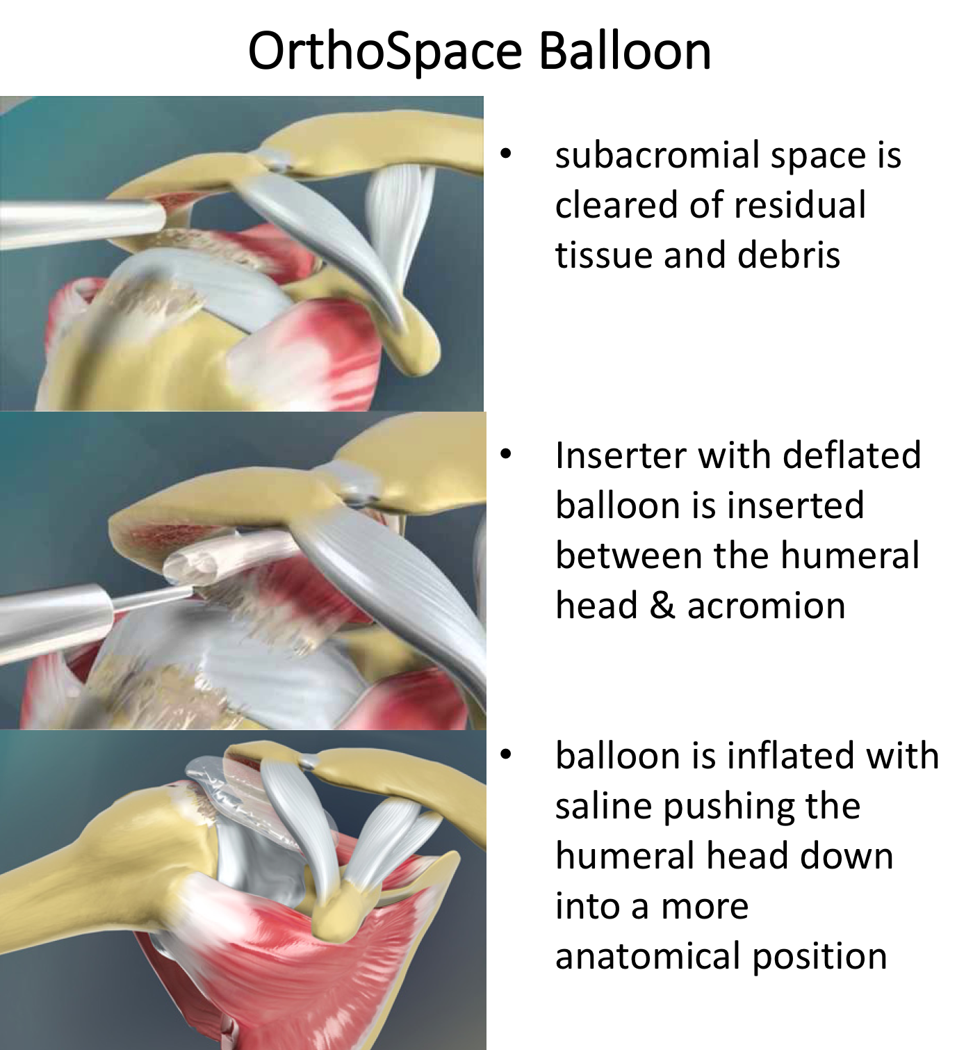
An Arthroscopic Orthospace Balloon procedure involves inserting an, initially deflated, balloon device into the space between the top of the humerus and undersurface of the acromion. The balloon is then gradually inflated with Normal Saline Fluid ‘pushing’ the humeral head downwards into its anatomical position. The balloon is then sealed off. Due to the shape of the undersurace of the acromion the balloon will be ‘jammed’ into position so that, as the Head of the Humerus and Shoulder move, the balloon remains in position. The balloon is mad of a ‘bio-absorbable’ material and will ‘disappear’ over the next 3 – 6 months.
The rational for the procedure is, that by re-poistioning the Humeral Head in its correct position, the accessory muscles around the shoulder have the opportunity to re-establish their normal working mechanism. At the same time they are, potentially, able to compensate for the deficient Rotator Cuff muscles.
An Arthroscopic Orthospace Balloon procedure is a relatively simple procedure with very few complications. Its success is variable with about 60 – 75% of patients obtaining a good result with respect to pain relief and a functional range of movement. However, it does not return the shoulder anatomy back to normal.
My routine Arthroscopic Orthospace Procedure is described below…
Watch a video of an arthroscopic Orthospace Balloon Insertion….
Arthroscopic Orthospace Balloon Procedure
- The patient is anaesthetised with a general anaesthetic and interscaelane nerve block
Find out more about having an anaesthetic…. (Patient Information – Having an Anaesthetic – General Anaesthetic)
Find out more about an Interscalene Nerve Block…. (Patient Information – Anaesthetic – Interscelene nerve block)
- A posterior, an anterior and a lateral portal are used to access the Gleno-Humeral Joint (Shoulder Joint) and Subacromial spaces
- The Glenohumeral Joint is initially viewed to assess the size and extent of the tear and to look for any other associated problems, particularly with regards to Osteoarthritic changes to the joint and problems with the Long Head of Biceps
- Using a Shaver and Radiofrequency wand the joint is then cleaned out removing any loose fragments and excising any ragged tissue
- If the Long Head of Biceps is considered to be damaged a Tenotomy is performed
- The Subacromial Space is then entered and the soft-tissue over the top of the Glenoid is cleared away, to create a ‘pocket’ into which the medial edge of the Orthospace balloon will be inserted
- The deflated Balloon is then inserted through the Lateral portal and its medial edge positioned in the pre-created ‘pocket’ above the Glenoid
- The Balloon is then inflated using Normal Saline solution. Inflation continues until the Humeral Head is considered to be in its correct position
- The shoulder is then moved through its full range of motion, to check that the Balloon is wedged securely
- At the end of the procedure the joint and subacromial space are assessed, washed out and the wounds closed with sub-cuticular sutures
After the Surgery
Post-Operative Care
Following an Arthroscopic Orthospace Balloon procedure the patient maybe able to go home on the same day as their surgery or may stay in the Hospital over-night. This would depend on various other factors. I would see the patient after the surgery to discuss how the procedure has gone and arrange for further Follow-Up. The patient will be seen by the In-Patient Physiotherapy team, who will instruct them on the initial Rehabilitation Protocol for their shoulder and will organise for further Out-Patient physiotherapy.
Find out more about Physiotherapy following Rotator Cuff Surgery….
I would usually review patients in the clinic 1 month and 3 months after their procedure to assess their progress and recovery.
Rehabilitation Protocol
Immediately after the surgery, when the patient has woken up from their general anaesthetic, their shoulder and arm will be numb from the interscalaene nerve block. This will usually last for 18 – 24 hours after the surgery. The patient’s arm will be in a Sling for this period to protect their arm. After the nerve block has worn off the arm can come out of the sling and can be used as normal.
After an Arthroscopic Orthospace Balloon Procedure, it is initially important to ‘protect’ the Balloon from any significant load and extreme movement. The physiotherapists will instruct the patient on the ‘safe zones’ of movement out of their sling over this period. Patients would usually need to have their arm in a sling for 2 weeks after the procedure. Occasionally, this can vary depending on technical factors and tissue quality related to the surgery.
My standard rehabilitation protocol is outlined below. The information and time to recovery are a general estimation and may vary from person to person.
|
Post op |
|
| Immediate |
|
| Day 1-3
Weeks |
|
| 3-6 Weeks |
|
|
Milestones |
|
|
Week 3 |
Full passive range of movement |
|
Week 6 |
Full active range of movement, good scapular control |
|
Return to Functional Activities |
|
| Driving |
|
| Swimming |
|
| Golf |
|
| Heavy Lifting |
|
| Work |
|
| Manual - as guided by surgeon | |
Success of Surgery, Risks & Complications
An Arthroscopic Orthospace Balloon procedure is a safe and reasonably reliable procedure with regards to alleviating pain and restoring variable function. Whilst some people notice a significant benefit within weeks of surgery it can take many months for others. Some patients describe a transient period of discomfort about 3 months after the procedure, that usually settles after a month. This may represent the Balloon beginning to ‘dissolve’. It is extremely rare that anyone’s shoulder is ever any worse than before the surgery.
There are always risks and complications associated with any operation.
- Anaesthetic - The risks of having a General Anaesthetic and an Interscalaene Nerve Block are very low, but will always need to be assessed on an individual basis by an Anaesthetist. Suffice it to say, that whilst a Shoulder Operation can in no way be considered a ‘life-saving’ procedure, an Anaesthetist would not consider undertaking an anaesthetic if they had any concerns that an undue risk was being taken
- Infection – Infection following arthroscopic surgery is rare < 0.2%
- Neurovascular Injury – Damage tosignificant neurovascular structures during arthroscopic shoulder surgery is rare < 0.2%
- CRPS Type 1 – A Chronic Pain Syndrome following arthroscopic shoulder surgery is rare < 0.2%
Outcome Measures
Assessing patient outcomes following surgery, using validated scoring systems, is a very important and useful exercise.
Calcific Tendonitis
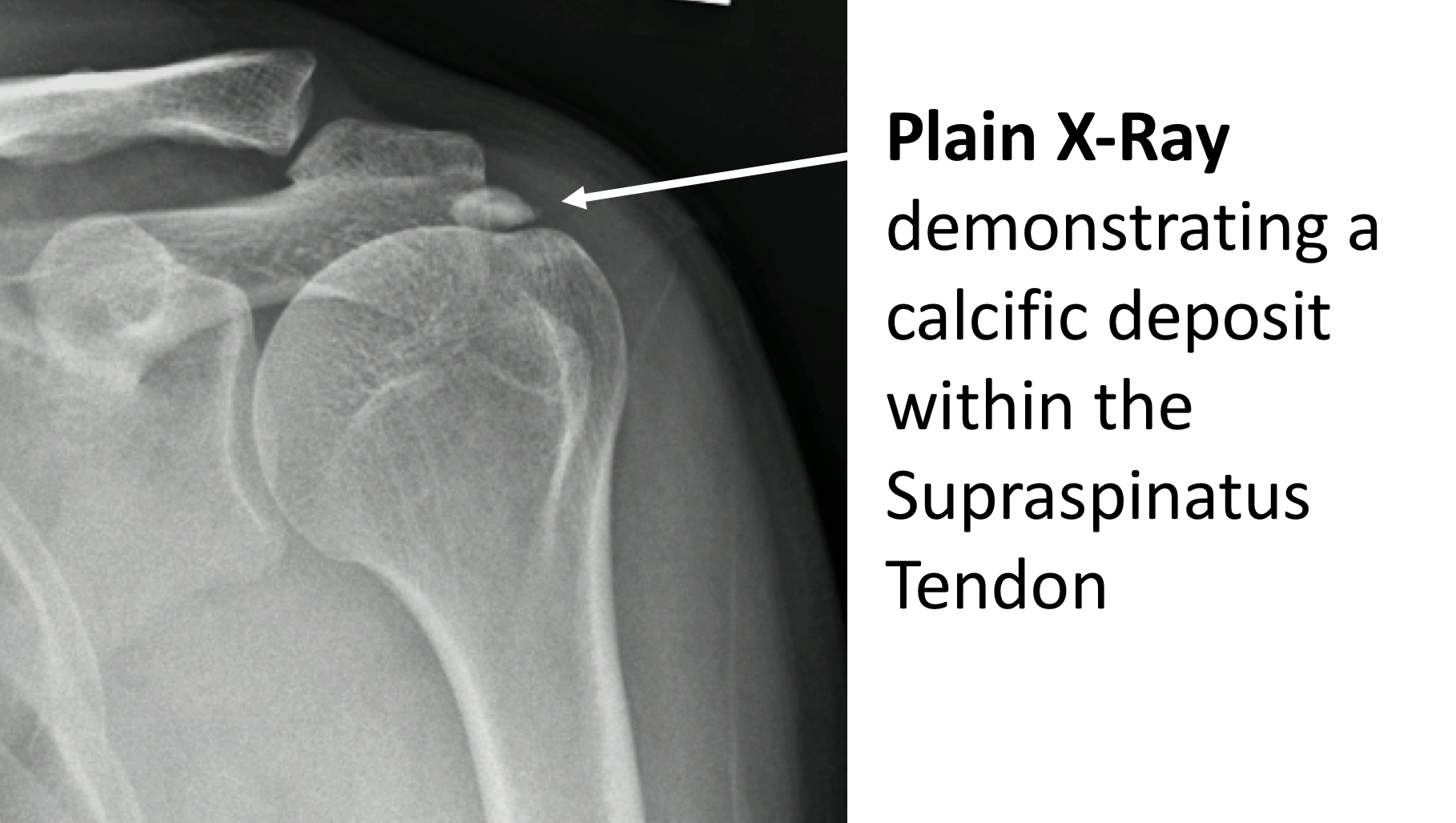
Calcific Tendonitis refers to calcium deposits forming in the rotator cuff tendons (most commonly the – Supraspinatus Tendon). Although, most of the time, calcific deposits are small and do not cause any pain, they can occasionally cause very significant problems. This can be due to a deposit causing pressure and mechanical symptoms and to chemical ‘irritation’.
There are 2 types of Calcific Tendonitis
Degenerative Calcific Tendonitis
- Degenerative Calcific Tendonitis can occur as part of normal degenerative wear and tear in rotator cuff tendons when some of the fibres begin to fray and wear. During the healing process calcium deposits are sometimes formed within the damaged tendons.
- This is often an ‘evolving process’ so that, over time, these calcium deposits form and are then slowly resorbed.
- this process is very common between the ages of 30 to 60 years, and is often painless, so that most of the time it is asymptomatic and people do not know that it is going on.
- calcific deposits can often be seen on plain x-rays, and it is estimated that 5% of the population between 30 to 60 years of age will have a calcium deposit on a shoulder x-ray, despite being asymptomatic.
- however, in certain people, the calcium deposit can become very large and lead to significant pain due to pressure within the tendon, impingement and chemical ‘irritation’.

Reactive Calcific Tendonitis
- Reactive Calcific Tendonitis is a different process and is not related to degeneration. The cause is not known, but it appears that, due metabolic changes within the tendon, calcium deposits are formed.
- As the deposits begin to form there can be a rapid increase in ‘pressure’ within the tendon, sometimes leading to an acute onset of severe pain
- Again, this is an ‘evolving’ process and over time the calcium deposits and are slowly resorbed.
- This process is relatively uncommon but can occur in people from the age of 20 to 60 years.
What are the Symptoms of Calcific Tendonitis
Chronic / Degenerative Calcific Tendonitis
- The symptoms of Chronic Calcific Tendonitis are identical to those of supraspinatus tendonitis or impingement
- In fact the cause of the pain is identical as essentially the enlarged and inflamed tendon impinges on the undersurface of the acromion
- As degenerative calcific deposits form slowly the onset of symptoms can often be quite insidious. As a result, many people find that Calcific Tendonitis gradually develops over time..
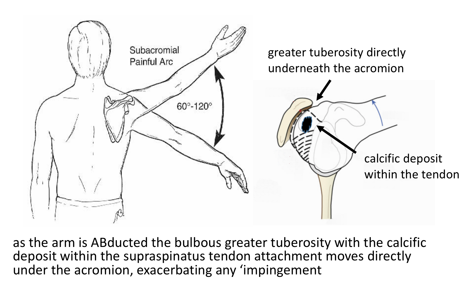
- There is a bulge on the side of the Humeral Head (Greater Tuberosity) where the Supraspinatus and Infraspinatus Tendons insert into the bone. As the shoulder moves, this bulge lies directly under the acromion between 60 and 120 degrees of elevation. This is the point where the space between the top of the Humeral Head and the Acromion is the narrowest. This is known as the ‘Painful Arc’ of movement. The early symptoms of Calcific Tendonitis tend to be a background pain over the shoulder with particular discomfort when elevating the shoulder within the ‘Painful Arc’. The shoulder is often uncomfortable to lie on at night.
- As symptoms deteriorate the shoulder becomes increasingly painful more of the time. Pain tends to occur on more directions of movement and the shoulder can appear to become weaker. Night discomfort often becomes more of a feature.
Acute / Reactive Tendonitis
- In Acute Calcific Tendonitis the calcific deposits usually form rapidly
- Symptoms can occur suddenly ‘out of the blue’ or maybe precipitated by a minor injury
- Sometimes, within a matter of hours, patients develop an ‘excruciating’ pain that is there constantly
- The pain can be so severe that even the strongest pain-killers do not seem to have an effect
How do you Diagnose Calcific Tendonitis?
- History – Most people notice a gradual onset of pain and discomfort over the top and side of their shoulder. Sometimes people can identify a specific injury or incident that triggered their problem. They may notice particular pain on elevating their arm and in bed at night. As the condition deteriorates they may get pain more of the time and on more directions of movement. They also might notice some weakness.
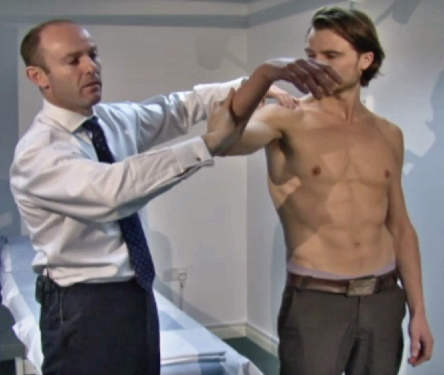
- Examination – Patients often experience some discomfort on palpation over the front edge of their Acromion directly over the insertion of the supraspinatus tendon, where the calcific deposit lies. People may have a ‘painful arc’ on elevating their shoulder and positive ‘impingement signs’. People with Supraspinatus tendonitis often have associated problems with Acromioclavicular Joint (ACjt) arthritis and Long Head of Biceps Tendonitis and may have symptoms and signs of these as well.
find out more about examination of the shoulder for rotator cuff disease… (Research & Education – Video – Examination of the Shoulder 3)
- Investigations – Chronic Calcific Tendonitis
- X-Ray –For Chronic Calcific Tendonitis an x-ray will usually demonstrate a calcific deposit sitting in the space underneath the acromion. This is where the supraspinatus tendon is situated.
find out more about x-rays…. (Patient Information – Imaging – X-Rays)
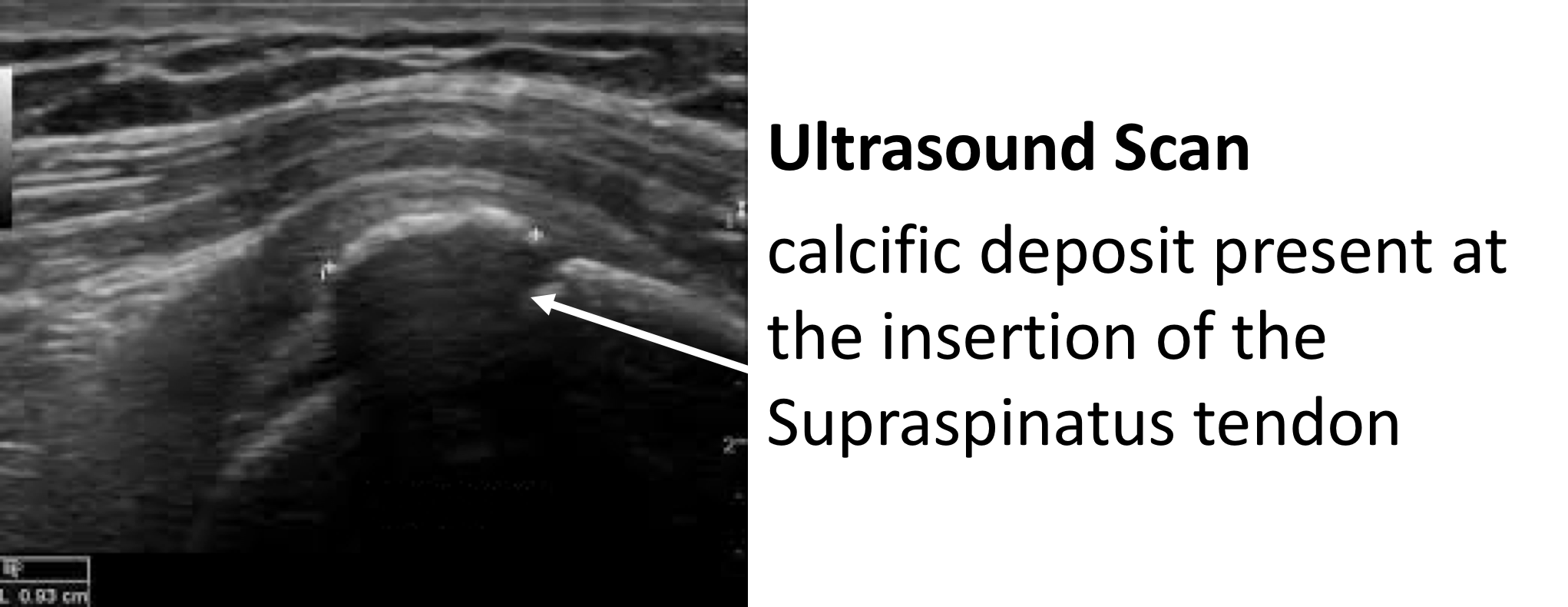
- Ultrasound Scan (USS) – An USS will nicely demonstrate the Rotator Cuff tendons and the Long Head of Biceps. It is able to show a Calcific Deposit and whether there is an associated tendonitis, partial or full thickness tear of the tendons.
find out more about USS….(Patient Information – Imaging – USS)
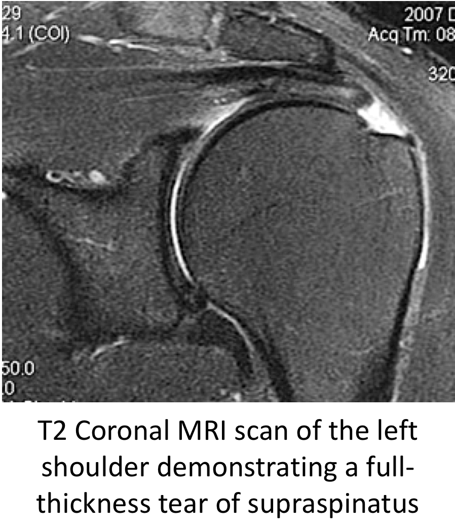
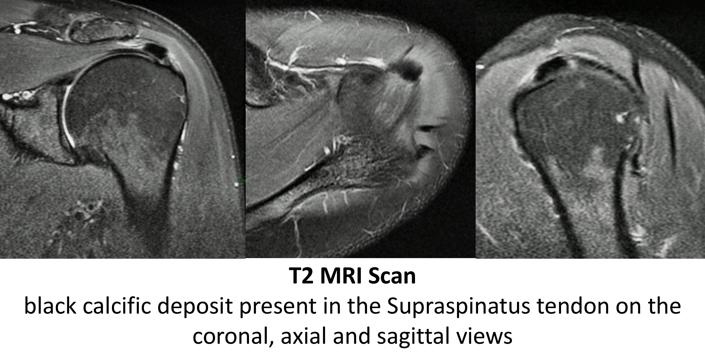
- MRI Scan- An MRI scan is the best investigation to visualize the rotator cuff. It can show all of the tendons and whether there is a calcific deposit, tendonitis and a partial or full thickness tear.
Find out more about MRI scans…. (Patient Information – Imaging – MRI)
- Investigations – Acute Calcific Tendonitis
- The symptoms of an Acute Calcific Tendonitis can develop so rapidly that insufficient calcium deposits may have been formed to be visualized on an X-Ray or an Ultrasound Scan in the early stages
- MRI Scan- An MRI scan is the best investigation to visualize the early changes associated with an Acute Calcific Tendonitis
Management of Chronic Calcific Tendonitis
The vast majority of patients with a Chronic Calcific Tendonitis can be successfully treated non-operatively with a combination of NSAIDs (Non-Steroidal Anti-Inflammatory Drugs) and Physiotherapy. The treatment is identical to that for Supraspinatus Tendonitis
- Physiotherapy – Physiotherapy and specific Rotator Cuff Strengthening exercises are the main-stay of the initial treatment for Chronic Calcific Tendonitis. By specifically strengthening the Supraspinatus muscle, where the deposit is usually located, it can work and contract more efficiently pulling the humeral head downwards and allowing the tendon to move more freely. If pain is a particular feature a short course of NSAIDs prescribed by a Physician can help.
find out more about Rotator Cuff Strengthening Exercises….(Patient Information – Physiotherapy – Physiotherapy-Rotator Cuff – Non-Operative)
- Analgesia/Pain Relief
- NSAID (Non-Steroidal Anti-Inflammatories) –
- NSAIDs work be reducing the painful inflammatory response
- NSAIDs can damage the stomach lining and affect the kidneys. It is important that a patient’s Family Doctor prescribes this medication if it is going to be used for a longer term
- Codeine based Analgesics –
- Codeine based analgesics are pain killers and affect a patient’s perception of pain. As a result, they can have some effect on consciousness depending on their strength
- Codeine based analgesics can lead to constipation if taken for a longer time. Having a high-fibre diet or even taking laxatives might need to be considered
- Nociceptive Analgesics –
- Nociceptive pain killers work on nerve generated pain
- Amitriptyline in lower doses works as a nociceptive pain killer. It has a useful side-effect in that it can make patients drowsy
- in cases of severe pain Amitriptyline can be prescribed at night
- NSAID (Non-Steroidal Anti-Inflammatories) –

- Subacromial Cortisone Injection –
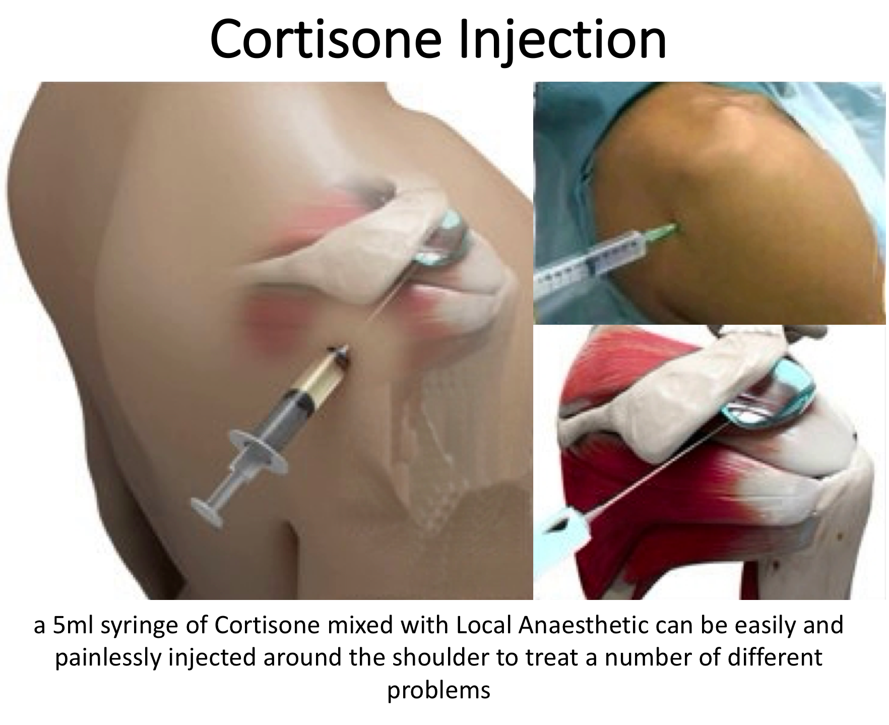
- If someone’s Chronic Calcific Tendonitis has failed to settle, after an adequate course of Physiotherapy, a Subacromial Cortisone injection can help to settle the symptoms down. Cortisone is a corticosteroid that is naturally produced by the body’s Adrenal Gland. Injectable Cortisone is synthetically produced and has a very powerful ant-inflammatory action. When injected into the Subacromial space it has the potential of settling severe inflammation, allowing the patient to undertake their rehabilitation exercises.
- A Subacromial Cortisone can be easily administered in the Out-Patient Clinic. It is a quick and relatively painless procedure. Afterwards patients can continue with their normal day-to-day activities. Occasionally patients can feel a bit of soreness around their shoulder later that day, but this usually passes fairly quickly
- It often takes several days before someone notices the benefits following a Cortisone injection and sometimes several weeks. The Cortisone works in the background and there is no specific requirement to particularly rest the shoulder or to do extra exercises. The full benefits of a Cortisone injection are usually felt within a month. In some cases, the Cortisone may not give any benefit, this may be an indication of the severity of the Supraspinatus Tendonitis.
- A Cortisone injection only lasts in the body for a few days. Any benefit that someone gets from the Cortisone will be from its acute anti-inflammatory effect allowing the Supraspinatus Tendonitis to settle. If the symptoms do return after a while, it is not because the Cortisone has worn off, but because the inflammation has returned.
- Infections can occur after any type of injection but are extremely rare (1 in 15,000). If someone feels that they are developing an infection within 48 hours of a Cortisone injection they should seek advice from their Family Practitioner.
- Surgery for Chronic Calcific Tendonitis – In some instances a Supraspinatus Tendonitis fails to respond, or continues to recur, despite adequate Physiotherapy and Cortisone treatment. In this situation the only treatment that is likely to settle the symptoms is Surgery.
Arthroscopic Subacromial Decompression, Excision of Calcific Deposit +/- Rotator Cuff Repair
The best operation for someone with a Chronic Calcific Tendonitis, that is refractory to treatment, is an Arthroscopic Subacromial Decompression and excision of the calcific deposit. This is an operation where an Arthroscopic camera is positioned inside the Subacromial Space and, by using specialized instruments, the undersurface of the front of the acromion is burred away. This creates more space for the Supraspinatus Tendon to run underneath the Acromion.
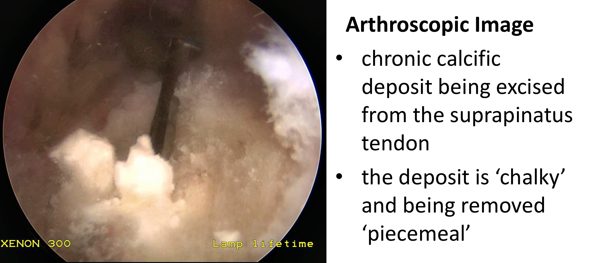
Following this the Calcific Deposit can be identified and removed. The deposit is usually of a chalky consistency within the tendon. So rather than being able to extract it as a single lump, it is usually the case of removing as many bits as possible. To do this, depending on the size of the deposit, may involve opening up the tendon which can result in a defect or hole being left behind. If this is the case a Rotator Cuff Repair is then undertaken.
An Arthroscopic Subacromial Decompression and Excision of Calcific deposit can usually be done as a Day Case procedure and the patient’s shoulder does not need to be immobilized for anytime afterwards, unless an additional rotator cuff repair has been undertaken. The surgery aims to remove the part of the Acromion and the calcific deposit that has been causing pain. It does not instantaneously deal with the underlying inflammation. However, by enabling the Supraspinatus Tendon to run freely and removing the deposit the symptoms can then begin to settle down. As a result it usually takes between 3 – 6 months to gain the full benefits of the procedure. It has a high success rate with around 95% of patients being happy with the result 6 months after their operation. My routine Arthroscopic Subacromial Decompression and Excision of Calcific Deposit Procedure is described below,
Watch a video of an Arthroscopic Subacromial Decompression… (Research & Education – Video – Subacromial Decompression)
Find out more about arthroscopic shoulder surgery….
- The patient is anaesthetised with a general anaesthetic and interscaelane nerve block
Find out more about having an anaesthetic….
Find out more about an Interscalene Nerve Block….
- A posterior and a single lateral portal are used for to view the Gleno-Humeral Joint (Shoulder Joint) and Subacromial spaces
- The Glenohumeral Joint is initially assessed to look for any other problems that might be associated with a Supraspinatus Tendonitis and, in particular, the Rotator Cuff and the Long Head of Biceps
- The Subacromial Space is then entered and the undersurface of the Acromion identified.
- A lateral portal is then created
- Using a combination of a powered shaver and radiofrequency probe the coracoacromial ligament is released, revealing the Subacromial Spur.
- A powered burr is then used to resect about 5mm of bone from the undersurface of the acromion
- The Decompression is then assessed to check that sufficient bone has been resected to alleviate the Impingment
- The Rotator Cuff is then examined and the Calcific Deposit identified
- Using a combination of a probe, punch and shaver the chalky Calcific Deposit is then removed
- After removal of the Calcific Deposit the Rotator Cuff Tendon is assessed and, if there is a significant defect, this will be then also be repaired
- The Subacromial Space is then washed out and the wounds closed with sub-cuticular sutures
After the Surgery
Post-Operative Care
Following an Arthroscopic Subacromial Decompression the patient is usually able to go home on the same day as their surgery. I would see the patient after the surgery to discuss how the procedure has gone and arrange for further Follow-Up. The patient will be seen by the In-Patient Physiotherapy team, who will instruct them on the initial Rehabilitation Protocol for their shoulder. Further Out-Patient physiotherapy will then be organised.
Find out more about Physiotherapy following an ASD….
I would usually review patients in the clinic 1 month and 3 months after their procedure to assess their progress and recovery.
Rehabilitation Protocol
Immediately after the surgery, when the patient has woken up from their general anaesthetic, their shoulder and arm will be numb from the interscalaene nerve block. This will usually last for 18 – 24 hours after the surgery. For this period, we advise patients to keep their arm in a sling, purely for protection. After the nerve block has worn off the arm can begin to be taken out of the sling. I encourage patients to wean themselves off of using the sling as quickly as possible, within the limits of discomfort.
My standard rehabilitation protocol is outlined below for a standard procedure WITHOUT an additional rotator cuff repair. The information and time to recovery are a general estimation and may vary from person to person.
|
Post op |
|
| Immediate |
|
|
Day 1-3 Weeks |
|
| 3-6 Weeks |
|
|
Milestones |
|
|
Week 3 |
Full passive range of movement |
|
Week 6 |
Full active range of movement, good scapular control |
|
Return to Functional Activities |
|
| Driving |
|
| Swimming |
|
| Golf |
|
| Racquet Sports/Repeated Overhead Activities |
|
| Lifting |
|
| Work |
|
Success of Surgery, Risks & Complications
An Arthroscopic Subacromial Decompression and Excision of Calcific Deposit is usually a very successful procedure but, because the surgery is only aimed at removing the spur of bone that has been impinging on the Supraspinatus Tendon, it usually takes between 3 – 6 months for the Tendonitis to settle and for the Shoulder to return to normal. For a standard Arthroscopic Subacromial Decompression>95% of patients’ will be happy with their shoulders after 6 months.
There are always risks and complications associated with any operation.
- Anaesthetic - The risks of having a General Anaesthetic and an Interscalaene Nerve Block are very low, but will always need to be assessed on an individual basis by an Anaesthetist. Suffice it to say, that whilst a Shoulder Operation can in no way be considered a ‘life-saving’ procedure, an Anaesthetist would not consider undertaking an anaesthetic if they had any concerns that an undue risk was being taken.
- Infection – Infection following arthroscopic surgery is rare < 0.2%
- Neurovascular Injury – Damage tosignificant neurovascular structures during arthroscopic shoulder surgery is rare < 0.2%
- CRPS Type 1 – A Chronic Pain Syndrome following arthroscopic shoulder surgery is rare < 0.2%
Outcome Measures
Assessing patient outcomes following surgery, using validated scoring systems, is a very important and useful exercise.
Management of Acute Calcific Tendonitis
- An Acute Calcific Tendonitis usually occurs so rapidly and is so painful that urgent treatment is required
- Subacromial Cortisone Injection –
- In some cases of an Acute Calcific Tendonitis a Cortisone injection can rapidly alleviate the pain and settle the symptoms down. Often within a few days
- Cortisone is a corticosteroid that is naturally produced by the body’s Adrenal Gland. Injectable Cortisone is synthetically produced and has a very powerful ant-inflammatory action. When injected into the Subacromial space it has the potential of settling the severe inflammation associated with an Acute Calcific Tendonitis, allowing the patient to undertake their rehabilitation exercises.
- A Subacromial Cortisone can be easily administered in the Out-Patient Clinic. It is a quick and relatively painless procedure. Afterwards patients can continue with their normal day-to-day activities. Occasionally patients can feel a bit of soreness around their shoulder later that day, but this usually passes fairly quickly.
- The Cortisone works in the background and there is no specific requirement to particularly rest the shoulder or to do extra exercises. The immediate benefits of a Cortisone injection should be felt within a few days. However, the full benefits of a Cortisone injection may take a month.
- Infections can occur after any type of injection but are extremely rare (1 in 15,000). If someone feels that they are developing an infection within 48 hours of a Cortisone injection they should seek advice from their Family Practitioner.
- Surgery for Acute Calcific Tendonitis – If the severe pain associated with an Acute Calcific Tendonitis does not begin to improve at all, within a few days, surgery is the only option to gain a rapid benefit.
Arthroscopic Excision of an Acute Calcific Tendonitis +/- Subacromial Decompression
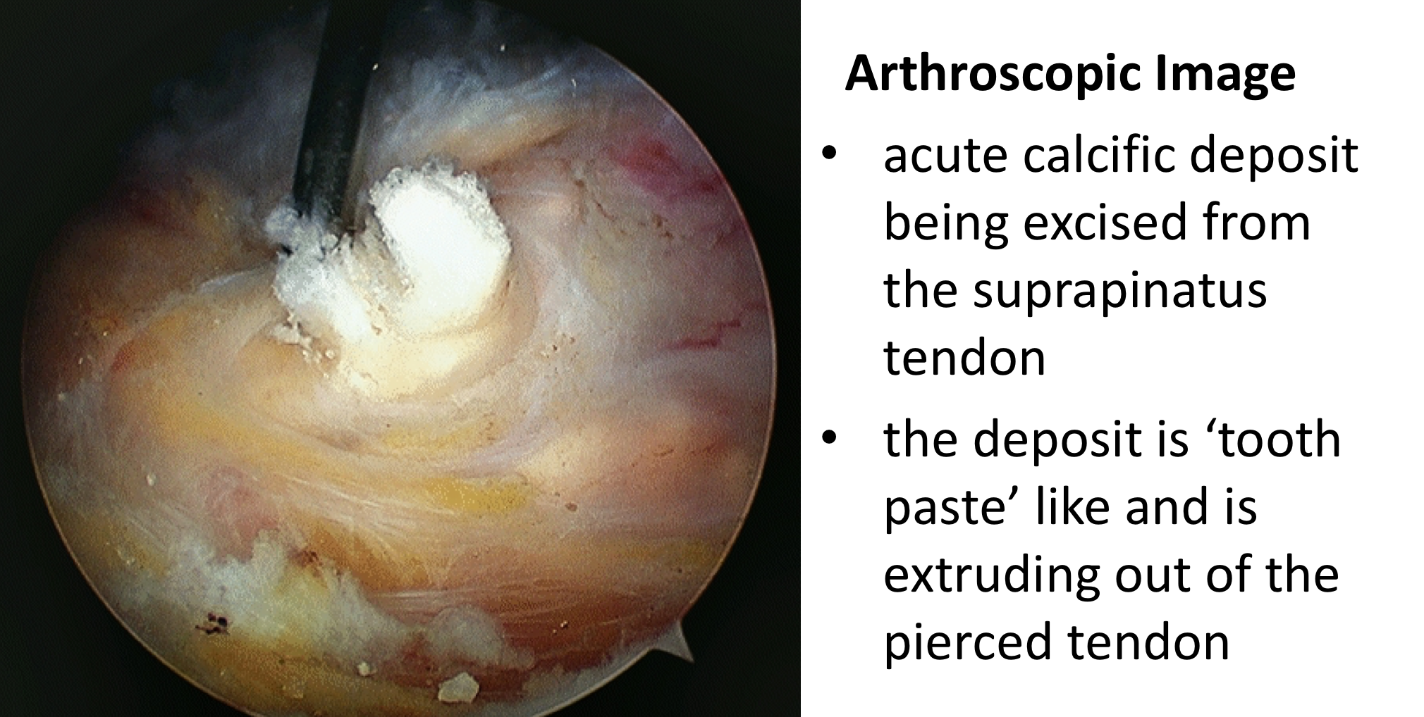
The best operation for someone with an Acute Calcific Tendonitis, that has not responded to a Cortisone injection, is an Arthroscopic Excision of the Acute Calcific deposit. This is an operation where an Arthroscopic camera is positioned inside the Subacromial Space and, by using specialized instruments, the Calcific Deposit is removed from within the tendon.
In an Acute Calcific Tendonitis the symptoms occur at such an early stage that the deposit is still usually in a fluid / ‘tooth-paste like’ stage. As a result, just opening up the tendon or multiple needle punctures are enough to allow it to fully extrude under pressure. The tendon does not need to be repaired and a decision can be made as to whether a Subacromial Decompression may help with recovery.
An Arthroscopic Excision of an Calcific deposit can usually be done as a Day Case procedure and the patient’s shoulder does not need to be immobilized for anytime afterwards, unless an additional rotator cuff repair has been undertaken. The surgery aims to extract the rapidly expanding deposit. It usually results in a major relief of the severe pain, but it can take between 3 – 6 months to gain the full benefits of the procedure. It has a high success rate with around 95% of patients being happy with the result 6 months after their operation. My routine Arthroscopic Excision of an Acute Calcific Deposit Procedure is described below,
Watch a video of an Arthroscopic Excision of an Acute Calcific Tendonitis… (Research & Education – Video – Acute Calcific Tendonitis)
- The patient is anaesthetised with a general anaesthetic and interscaelane nerve block
Find out more about having an anaesthetic….
Find out more about an Interscalene Nerve Block….
- A posterior and a single lateral portal are used for to view the Gleno-Humeral Joint (Shoulder Joint) and Subacromial spaces
- The Glenohumeral Joint is initially assessed to look for any other problems that might be associated with a Supraspinatus Tendonitis and, in particular, the Rotator Cuff and the Long Head of Biceps
- The Subacromial Space is then entered and the undersurface of the Acromion identified.
- A lateral portal is then created
- The Supraspinatus Tendon is then inspected and the area of the Calcific Deposit identified
- Either using a needle or a punch the tendon is pierced and the Calcific Deposit seen to extrude, rather like ‘tooth-paste’ coming out of a tube
- After removal of the Calcific Deposit the undersurface of the anterior acromion is assessed and, if there is a significant prominence, a decompression performed
- The Subacromial Space is then washed out and the wounds closed with sub-cuticular sutures
After the Surgery
Post-Operative Care
Following an Arthroscopic Excision of an Acute Calcific Deposit the patient is usually able to go home on the same day as their surgery. I would see the patient after the surgery to discuss how the procedure has gone and arrange for further Follow-Up. The patient will be seen by the In-Patient Physiotherapy team, who will instruct them on the initial Rehabilitation Protocol for their shoulder. Further Out-Patient physiotherapy will then be organised.
Find out more about Physiotherapy following an ASD….
I would usually review patients in the clinic 1 month and 3 months after their procedure to assess their progress and recovery.
Rehabilitation Protocol
Immediately after the surgery, when the patient has woken up from their general anaesthetic, their shoulder and arm will be numb from the interscalaene nerve block. This will usually last for 18 – 24 hours after the surgery. For this period, we advise patients to keep their arm in a sling, purely for protection. After the nerve block has worn off the arm can begin to be taken out of the sling. I encourage patients to wean themselves off of using the sling as quickly as possible, within the limits of discomfort.
My standard rehabilitation protocol is outlined below for a standard procedure. The information and time to recovery are a general estimation and may vary from person to person.
|
Post op |
|
| Immediate |
|
|
Day 1-3 Weeks |
|
| 3-6 Weeks |
|
|
Milestones |
|
|
Week 3 |
Full passive range of movement |
|
Week 6 |
Full active range of movement, good scapular control |
|
Return to Functional Activities |
|
| Driving |
|
| Swimming |
|
| Golf |
|
| Racquet Sports/Repeated Overhead Activities |
|
| Lifting |
|
| Work |
|
Success of Surgery, Risks & Complications
An Arthroscopic Subacromial Excision of an Acute Calcific Deposit is usually a very successful procedure but, because the shoulder and tendon have been very inflamed it can take between 3 – 6 months for the Shoulder to return to normal. For a standard Arthroscopic Subacromial Decompression>95% of patients’ will be happy with their shoulders after 6 months.
There are always risks and complications associated with any operation.
- Anaesthetic - The risks of having a General Anaesthetic and an Interscalaene Nerve Block are very low, but will always need to be assessed on an individual basis by an Anaesthetist. Suffice it to say, that whilst a Shoulder Operation can in no way be considered a ‘life-saving’ procedure, an Anaesthetist would not consider undertaking an anaesthetic if they had any concerns that an undue risk was being taken.
- Infection – Infection following arthroscopic surgery is rare < 0.2%
- Neurovascular Injury – Damage tosignificant neurovascular structures during arthroscopic shoulder surgery is rare < 0.2%
- CRPS Type 1 – A Chronic Pain Syndrome following arthroscopic shoulder surgery is rare < 0.2%
Outcome Measures
Assessing patient outcomes following surgery, using validated scoring systems, is a very important and useful exercise.